
|
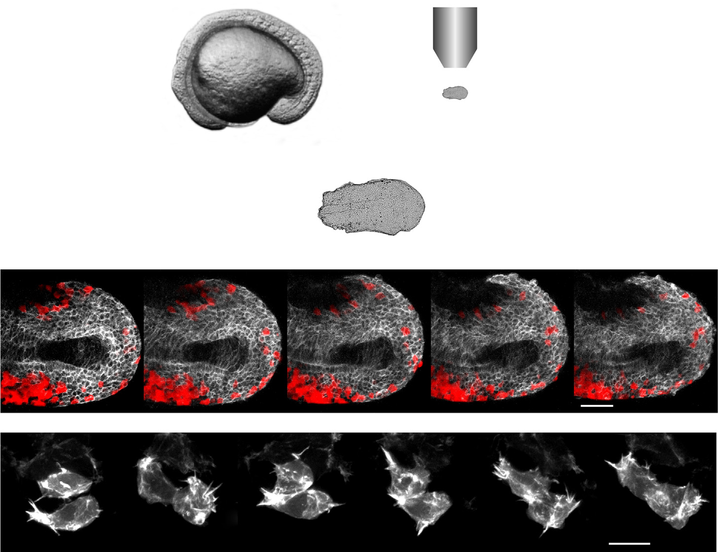
Fig. 2
A novel tailbud explant method allows for high spatio-temporal imaging of migrating cells in vivo. (A) The posterior portion of an embryo in mid-somitogenesis is dissected away from the anterior tissue and yolk, mounted, and imaged. (B) Time lapse image series of an explant from a wild-type host expressing a fluorescent membrane marker driven by the tbx16 promoter (white) with wild-type donor cells (red) taken at mid-somitogenesis. Anterior is to the left with the notochord running down the middle of the tissue. These images correspond to frames from Movie S1. Scale bar=50 µm. (C) Time lapse image series of two cells in an explant from a wild-type embryo mosaically expressing the fluorescent actin marker LifeAct driven by the tbx16 promoter. Scale bar=10 µm.
Reprinted from Developmental Biology, 406(2), Manning, A.J., Kimelman, D., Tbx16 and Msgn1 are required to establish directional cell migration of zebrafish mesodermal progenitors, 172-85, Copyright (2015) with permission from Elsevier. Full text @ Dev. Biol.