
|
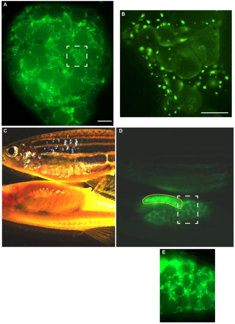
Fig. S2 Expression of Buc-GFP in ovaries. (A, B) Living ovary from an adult F1- female transgenic for Buc-GFP. Dashed square highlights the magnified area in B. Scale bar: 700 µm. (B) Magnification showing that the fluorescence of Buc-GFP is obscured during oogenesis probably by the uptake of yolk protein. Scale bar: 200 µm. (C-E) Comparison of a wild-type (top) and casper female (bottom) carrying a Buc-GFP transgene in incident (C) and fluorescent light (D). Yellow line encompasses the stomach with autofluorescent food residues in the digestive tract. Dashed square highlights the magnified area in (E).
Reprinted from Gene expression patterns : GEP, 18(1-2), Riemer, S., Bontems, F., Krishnakumar, P., Gömann, J., Dosch, R., A functional Bucky ball-GFP transgene visualizes germ plasm in living zebrafish, 44-52, Copyright (2015) with permission from Elsevier. Full text @ Gene Expr. Patterns