
|
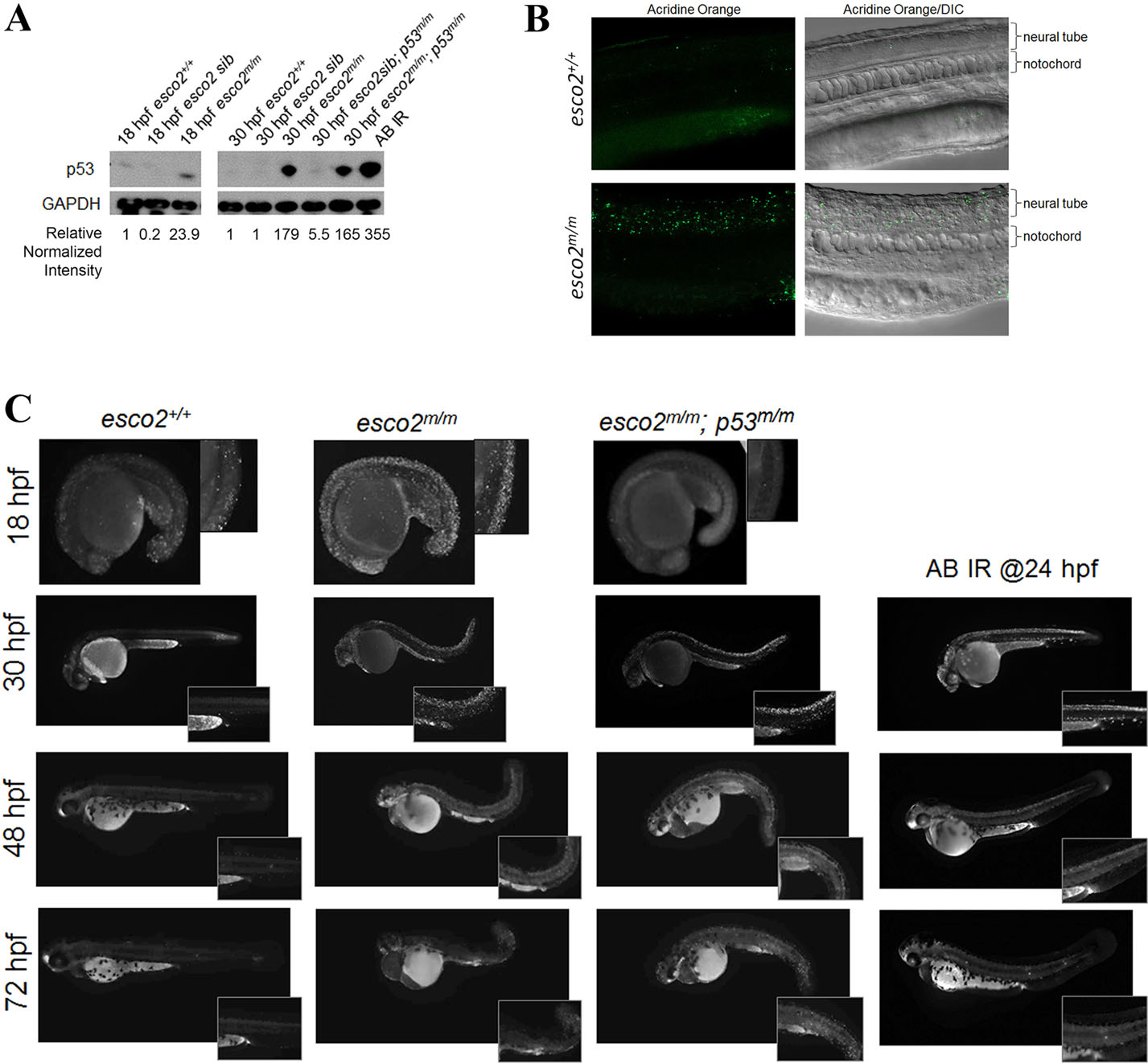
Fig. 2
p53 activation and neural tube apoptosis is an early consequence of loss of esco2. (A) Western blot for p53 protein levels in protein extracts from AB (esco2+/+), esco2 sib (esco2+/+ and esco2+/m), esco2 mutant (esco2m/m), esco2sib; p53m/m and esco2m/m; p53m/m embryos at 18 and 30hpf. Irradiated (IR) embryos at 100Gy were used as a positive control. Relative intensities were determined using ImageJ; each sample was normalized to GAPDH intensity, and then relative expression was calculated against esco2+/+ (relative normalized intensity=1). (B) Fluorescent and DIC/fluorescent merge images of esco2+/+ and esco2m/m 24-hpf embryos stained with acridine orange. (C) Acridine orange time-course staining spatially displaying apoptotic cells in esco2+/+, esco2m/m and esco2m/m; p53m/m. AB embryos irradiated at 24hpf were used as a positive control for DNA-damage-induced neural tube apoptosis. Insets depict higher magnification to visualize neural tube apoptosis.