Image
Figure Caption
Fig. S4
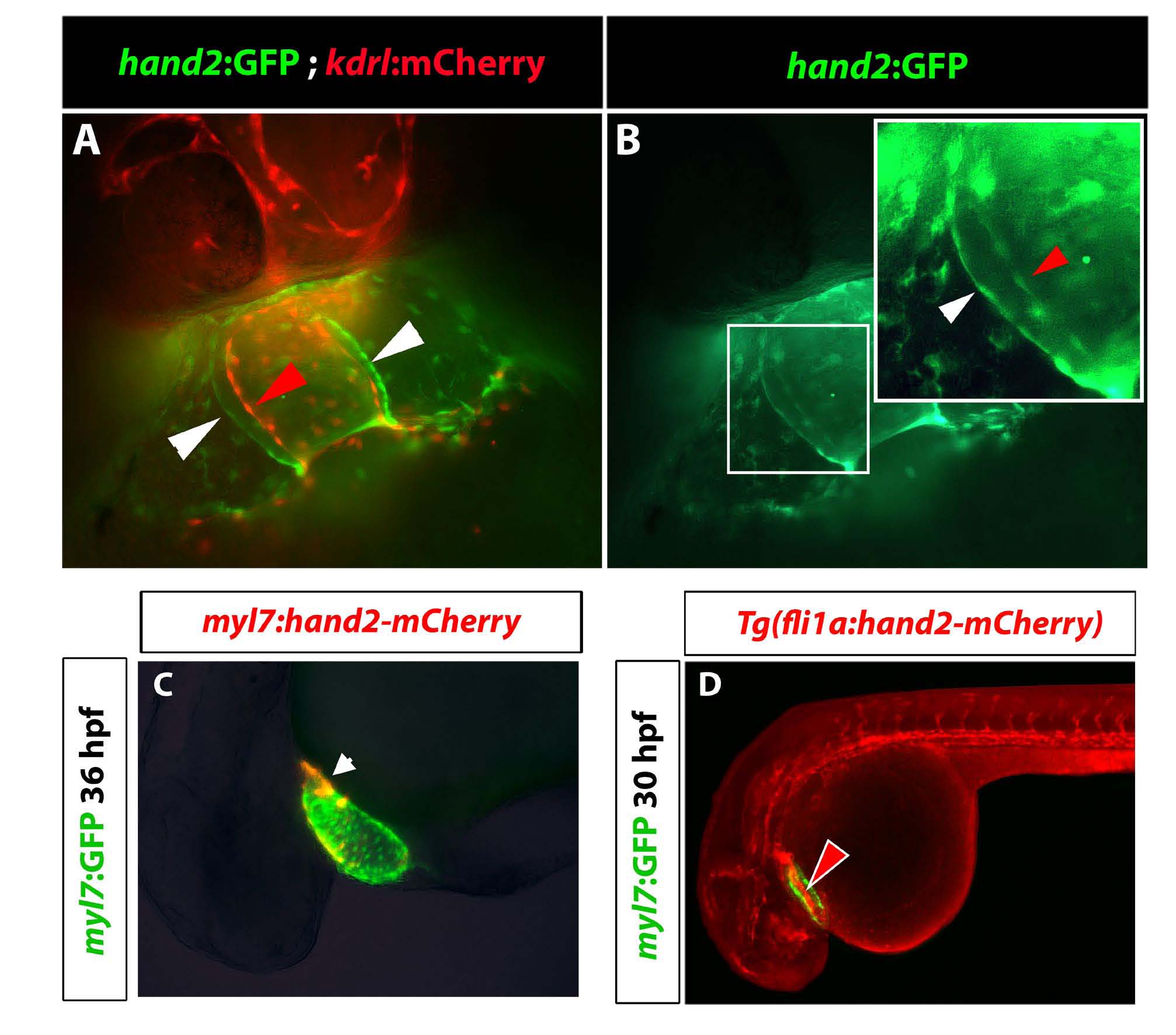
Hand2:GFP, myl7:hand2-mCherry and fli1a:hand2-mCherry reporter expression.
(A,B) Fluorescent imaging of cardiac region in hand2:GFP; kdrl:mCherry transgenic embryos at 35 hpf. (A) kdrl:mCherry+ endocardial cells are shown in red and hand2:GFP+ myocardial cells in green. (B) GFP channel shows both myocardial (white arrowhead) and endocardial cells (red arrowhead) labeled with hand2:GFP. (C) Mosaic expression of injected myl7:hand2-mCherry construct overlaps with Tg(myl7:GFP) fluorescence at 36 hpf. Fluorescence in lightly fixed embryos is shown. (D) mCherry fluorescence is observed in the vasculature and the endocardium at 30 hpf in lightly fixed Tg(fli1a:hand2-mCherry); Tg(myl7:GFP) embryos.
Acknowledgments
This image is the copyrighted work of the attributed author or publisher, and
ZFIN has permission only to display this image to its users.
Additional permissions should be obtained from the applicable author or publisher of the image.
Full text @ Development

