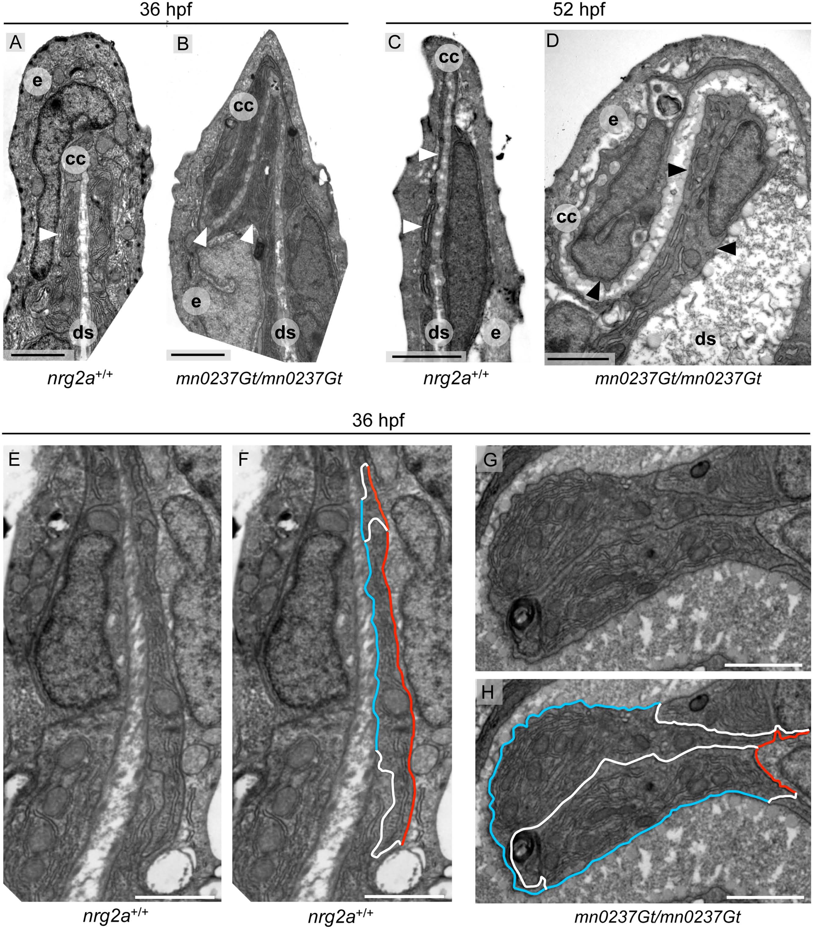
Fig. 8 MFF ridge cell in nrg2a mutants display alterations in basolateral versus apical dimensions.
(A-H) Transmission electron micrographs (TEM) of the distal-most region within wild-type and nrg2a mutant MFFs at 36 hpf (A, B, E-H) or 52 hpf (C,D). (A) By 36 hpf, wild-type ridge cells (arrowhead) have begun elongating laterally. Their apical and basal surfaces are parallel to each other and to the dermal space. (C) By 52 hpf, wild-ridge cells have continued elongating and have maintained their arrangements. (B, D) In nrg2a mutants (mn0237Gt/mn0237Gt), ridge cells (arrowheads) are morphologically distorted, fail to stay aligned parallel to the direction of the fin, and bulge into the dermal space, giving it a serpentine-like appearance. (E-H) Relative basolateral and apical dimensions in nrg2a mutants are distorted compared to their wild-type counterparts. To illustrate the changes, identical images are shown side by side with and without marked ridge cell borders. (E, F) By 36 hpf, apical (red) and basal (blue) borders of wild-type ridge cells are roughly parallel and of comparable lengths; lateral borders with neighboring basal keratinocytes are in white. (G, H) An example of mn0237Gt/mn0237Gt mutant ridge cells bulging into the dermal space. The pictured bulge consists of two adjacent ridge cells sharing an exaggeratedly lengthened lateral border (white) and with enlarged basal (blue) borders, but strongly reduced apical borders (red). Scale bars: 2 µm. Abbreviations: ds, dermal space; e, EV; cc, cleft cell; arrowheads point to ridge cells.

