
|
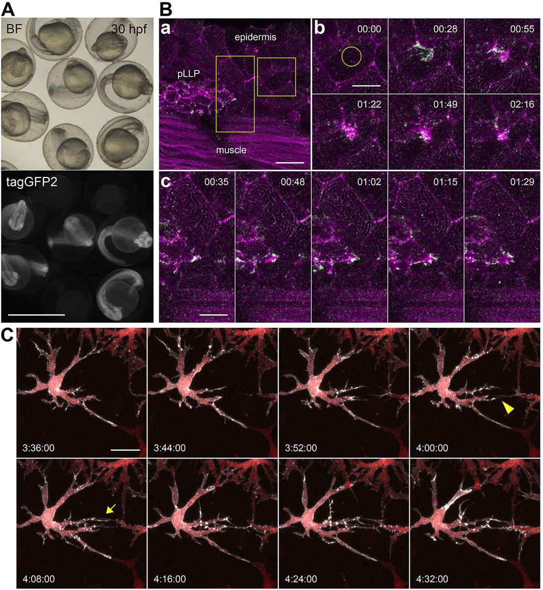
Fig. 2
Actin-CB-expressing transgenic zebrafish embryos reveal fast actin dynamics in multiple cell types. (A) 30-hpf embryos from an outcross of hsp70l:Actin-CB founder fish. The embryos were heat-shocked at 24hpf. Chromobody-expressing individuals show widespread fluorescence and no morphological abnormalities when compared with non-transgenic siblings. BF, brightfield. (B) Actin-CB can trace fast protein dynamics in living zebrafish. (a) Overview of a 36hpf embryo, in which actin-CB expression was induced by heat-shock at 24hpf. pLLP cells are visible, together with epidermal cells and muscle fibres. (b) Reorganization of actin after laser-induced wounding (yellow circle). (c) Detailed actin activity at the leading edge of front cells during pLLP migration. Magenta indicates signal from time frame t+1. Green indicates signal obtained from the subtraction of frame t from frame t+1. Green signal highlights the appearance of actin-CB signal from frame to frame (scanning interval is 7s). (C) Filopodial activity during establishment of intercellular contacts between neighbouring xanthophores. Detail of a csf1ra:gal4 embryo expressing UAS:NTR-mcherry (red) and UAS:Actin-CB (white). Filopodia extruded from xanthophore branches are directional to the prospective contact site with neighbouring cells (arrowhead and arrow). Time-stamps: min:s (B), h:min:s (C). Scale bars: 1mm in A; 10µm in Bb,c; 20µm in Ba,C.