
|
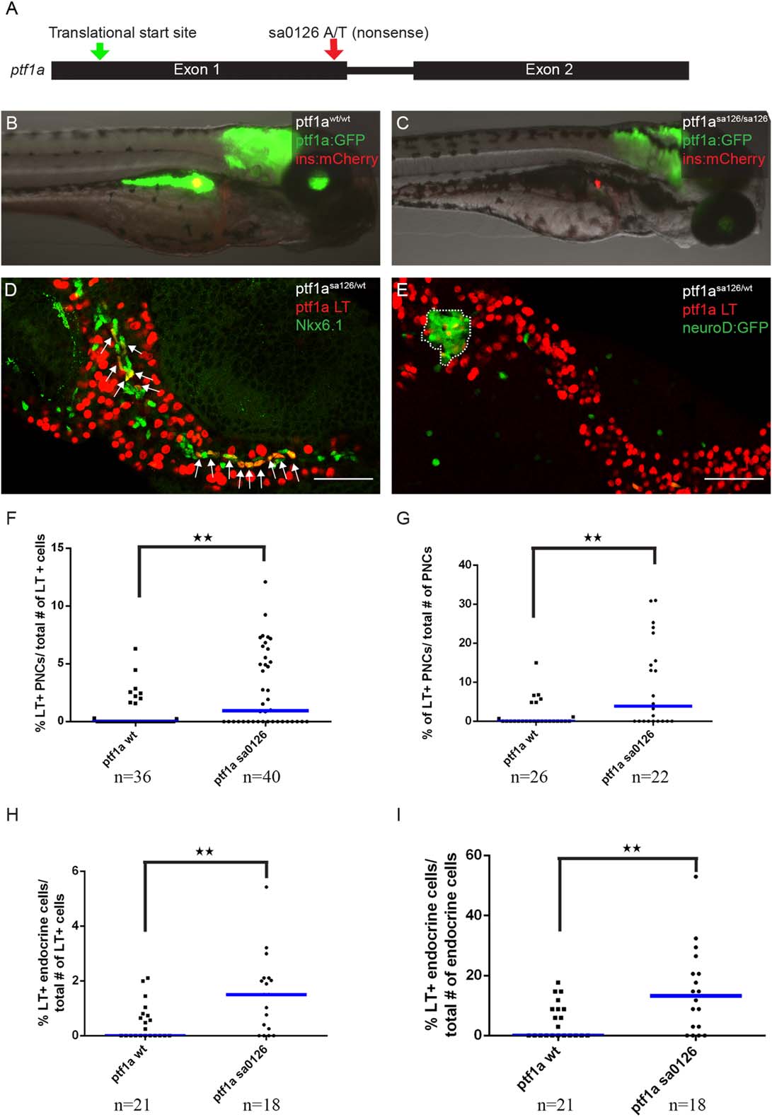
Fig. 5
Ptf1a haploinsufficiency increases the allocation of pancreatic progenitor cells to non-acinar lineages. A: The ptf1asa126 allele. This allele has an A to T transition in the first exon of ptf1a coding sequence, introducing a premature stop codon. B: Wildtype larvae, with ptf1a:GFP (green) marking acinar cells and ins:mCherry (red) transgene marking β cells. C: ptf1asa126/sa126 homozygous fish do not have exocrine pancreas. D, E: The ptf1a lineage is labeled by nuclear-mCherry expression (red). D: In ptf1asa125/wt fish, more lineage-labeled cells can be traced into Nkx6.1+ (green) PNCs. Arrows point to co-labeling events. E: In pf1asa126/wt fish, more lineage-labeled cells can be traced into the neuroD:GFP+ endocrine compartment. The principal islet is outlined. F, G: Quantification of ptf1a lineage-labeled cells contributing to PNCs, normalized by the total number of ptf1a-lineage labeled cells (F), or by the total number of PNCs (G). H, I: Quantification of ptf1a lineage–labeled cells contributing to the endocrine compartment, normalized by total number of ptf1a lineage–labeled cells (H), or by the total number of endocrine cells (I). F–I: “n” indicates the number of fish quantified for each genotype. Blue bars show population median.