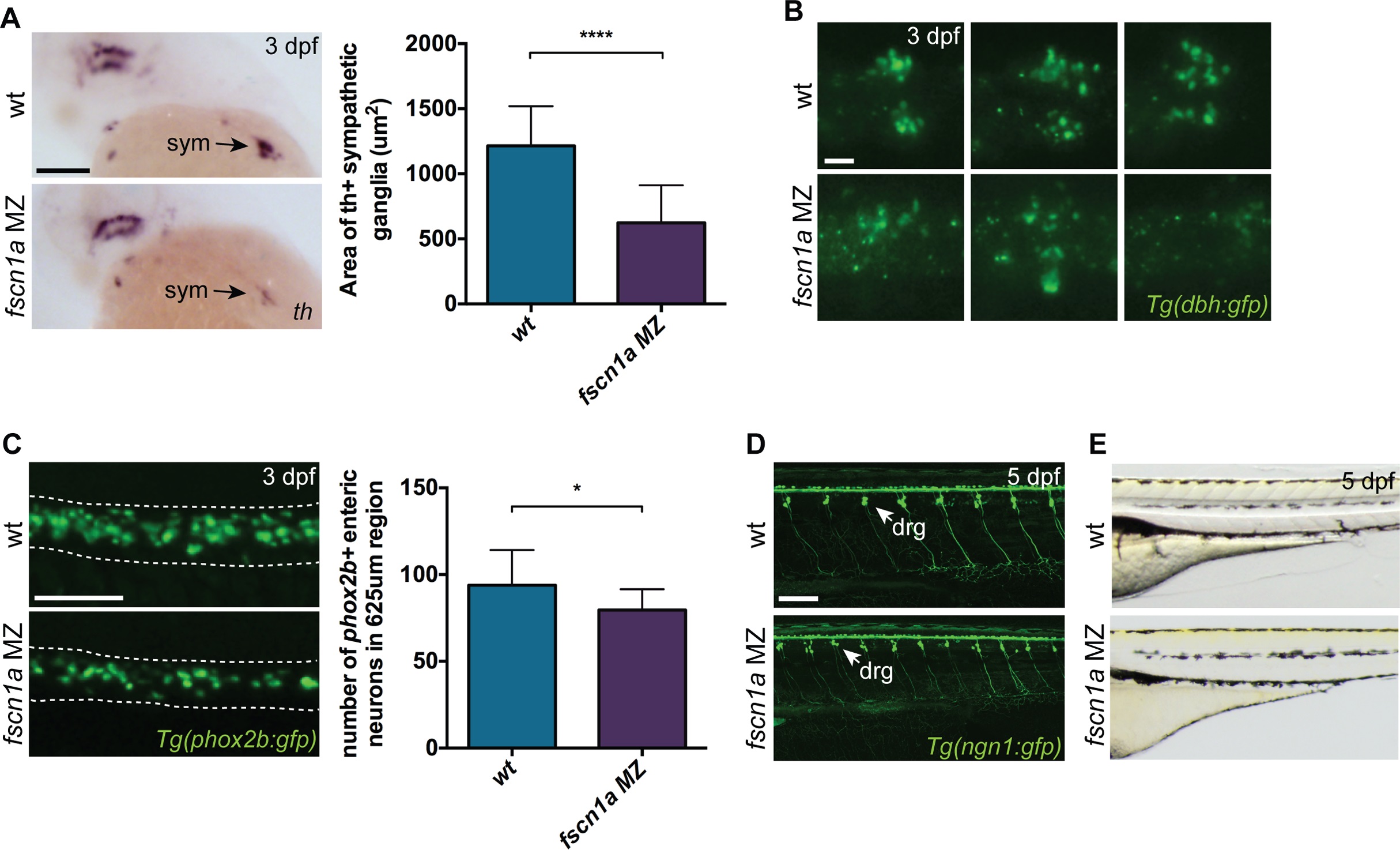
Fig. 7 fscn1a is required for development of a subset of NC derivatives.
(A) Lateral views of 3 dpf wt and fscn1a MZ embryos stained for th. sym = sympathetic ganglia, scale bar = 100 µm. Mean area of th+ sympathetic ganglia is plotted in graph on right. (n = 25 wt, 24 fscn1a MZ, ****p<0.0001). (B) Representative images of dbh:gfp+ sympathetic ganglia in 3 dpf wt and fscn1a MZ embryos. Dorsal views, anterior to the left. Scale bar = 25 µm. (C) Representative images of phox2b:gfp+ enteric neurons in 3 dpf wt and fscn1a MZ embryos. Dotted lines outline gut. Scale bar = 100 µm. Number of phox2b:gfp+ enteric neurons in 625 µm region of gut is plotted in graph on right (n = 15 wt, 13 fscn1 MZ, *p = 0.03). (D) ngn1:gfp+ dorsal root ganglia (drg) in 5 dpf wt and fscn1a MZ embryos. Scale bar = 100 µm. (E) Lateral views showing melanophores in 5 dpf wt and fscn1a MZ embryos.

