Fig. 1
Detection of Chitin in Zebrafish and Axolotl
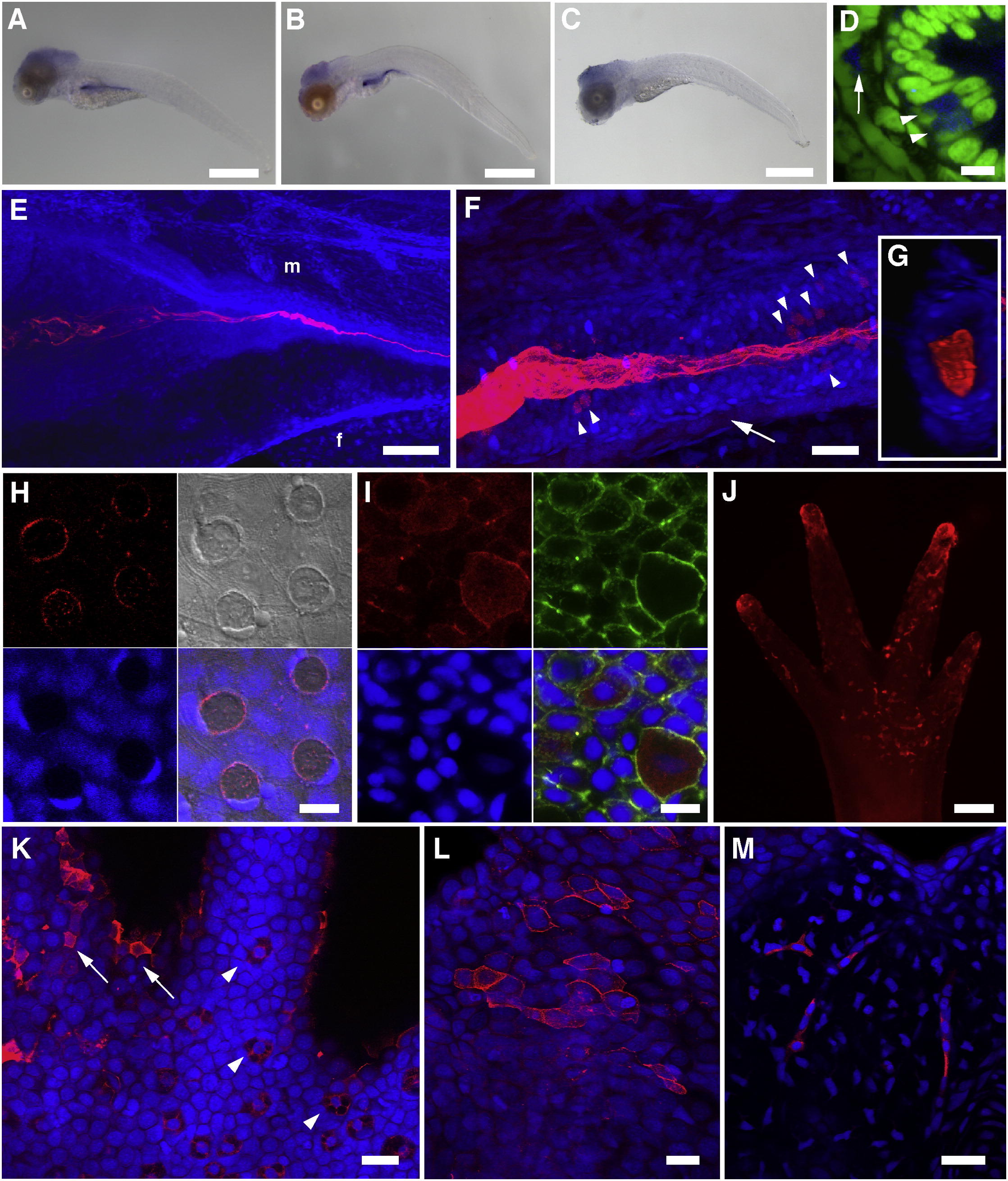
(A–D) In situ hybridization for chs1. NBT/BCIP signals were observed primarily in the developing digestive tract in 5 dpf (A) and 10 dpf (B) larvae, indicating that chs1 is expressed during larval development and in the gut. A representative “sense” control larva is shown in (C). No NBT/BCIP signals were observed in the developing gut of control larvae at either 5 or 10 dpf, although background staining was observed in the cranial region. Scale bars represent 500 µm.
(D) The ventral region of the mid-intestine of a 10 dpf larva (transverse section, different specimen than B) shows chs1 expression in select cells within the columnar intestinal epithelium (arrowheads) and in mesenchymal cells adjacent to the intestinal epithelium (arrow). NBT/BCIP deposition was imaged in the infrared range with confocal microscopy (blue), and nuclei were stained with Sytox Green (green). Scale bar represents 10 µm.
(E–M) A chitin probe (CBD-546, shown in red) was used for affinity histochemistry on fixed whole-mount specimens of zebrafish larvae (E–G) and scales (H and I) and axolotl larval forelimbs and tails (J–M). Nuclei were stained with TOTO-3 (blue), and rostral is to the left in (E) and (F).
(E) 3D-rendered projection of a 4 dpf zebrafish larva. “f” and “m” represent preanal finfold and somitic muscle, respectively; the “m” designation is above the approximate junction of the middle intestine. Chitin is observed in the developing larval gut, appearing fibrillar in the lumen of the intestinal bulb and coalescing as the lumen narrows posteriorly. Scale bar represents 100 µm.
(F) 3D-rendered projection of the mid-intestinal lumen from a 5 dpf zebrafish larva. The fibrous nature of the chitin is obvious, and several cells within the intestinal epithelial wall are observed that show strong chitin signals (arrowheads). A population of mesenchymal cells was also found to be positive for chitin signals (arrow). Scale bar represents 20 µm.
(G) Transverse section from a 3D volume-rendered reconstruction of the image in (F) showing chitin distributed throughout the lumen of the mid-intestine.
(H) Confocal image of a field of zebrafish scale epithelium. Panels show the following: CBD-546 (top left), DIC (top right), Sytox Green nuclear counterstain (pseudocolored in blue, bottom left), and merged image (bottom right). The four circular structures with red chitin signals are goblet cells that show polarized, crescent-shaped nuclei; polygonal concentric structures on the DIC overlay are squamous epithelial cells. Aggregations of small red punctate spots probably represent extracellular chitin. Scale bar represents 10 µm.
(I) Maximum intensity projection of a field of scale epithelial cells in which chitin is prominent throughout the cytoplasm of club cells. Panels show the following: CBD-546 (top left), phalloidin-FITC (top right), TOTO-3 nuclear counterstain (bottom left), and merged image (bottom right). The scale specimen was simultaneously stained for F-actin using phalloidin-FITC to help distinguish individual cells. Scale bar represents 10 µm.
(J) A forelimb (palm side up) of a larval axolotl after chitin histochemistry, imaged on a stereoscope. The tips of the developing digits and the palm area that show extensive chitin signals are primarily superficial epithelial cells. Scale bar represents 200 µm.
(K) A field from the base of a digit from a different forelimb than shown in (J), imaged by confocal microscopy. Arrowheads indicate several specialized Leydig epidermal cells, and the arrows identify a few of the superficial epithelial cells seen in (J). Scale bar represents 50 µm.
(L and M) Caudal fin of larval axolotl.
(L) Maximum intensity projection showing a subset of surface epithelial cells that exhibit strong chitin signals. Scale bar represents 50 µm.
(M) A deeper optical section showing strong chitin signals in the fibroblast-like cells of the fin mesenchyme. Scale bar represents 50 µm.
See also Figures S1 and S2.

