
|
Fig. 2
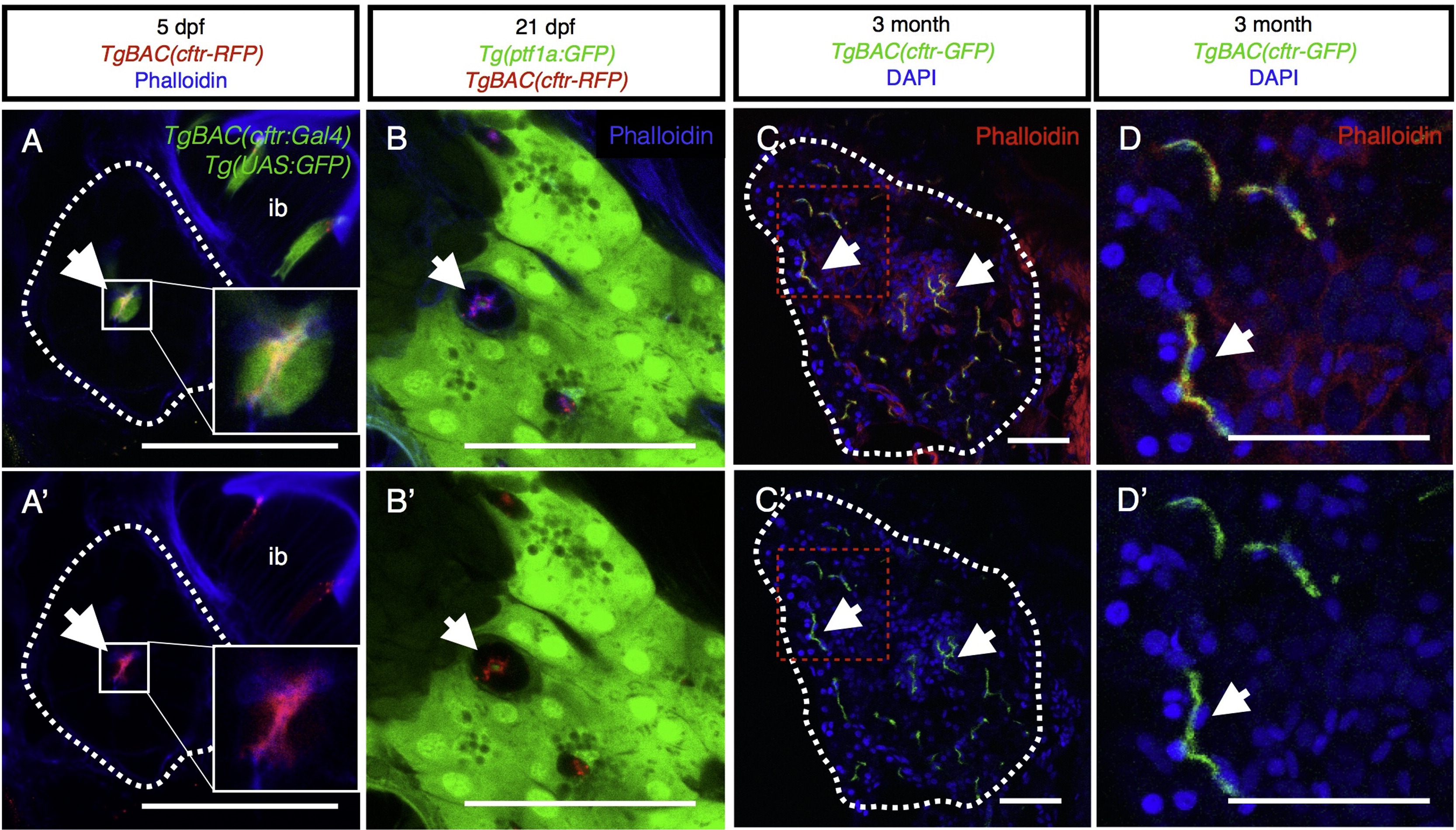
Cftr is localized to the apical membrane of the pancreatic duct epithelium throughout life. (A and A′) Transverse section of 5 dpf larvae expressing TgBAC(cftr-RFP) to show Cftr-RFP localization and TgBAC(cftr:Gal4); Tg(UAS:GFP) to mark the cytosol of cftr expressing cells with GFP. Dashed white lines indicate the periphery of pancreas. Inset is 3× magnification of ductal cells. ib: intestinal bulb. (B and B′) Transverse section of a 21 dpf larvae demonstrating Cftr-RFP localization at the apical membrane of the pancreatic duct in conjunction with TgBAC(ptf1a-GFP) expressed in the acinar cells. Cftr is observed at or near the apical membrane marked with phalloidin. (C and C2) Transverse section of 3 month, adult pancreas indicating Cftr-GFP expression in pancreatic ducts throughout the pancreas. Dashed white lines indicate the periphery of the pancreas. Red dashed box indicates inset (D). (D and D′) Cftr-GFP is localized at or near the apical membrane of the pancreatic ducts, marked by phalloidin staining. Arrows indicate ductal expression of Cftr. Scale bars=50 µm.
Reprinted from Developmental Biology, 399(2), Navis, A., Bagnat, M., Loss of cftr function leads to pancreatic destruction in larval zebrafish, 237-48, Copyright (2015) with permission from Elsevier. Full text @ Dev. Biol.