
|
Fig. S1
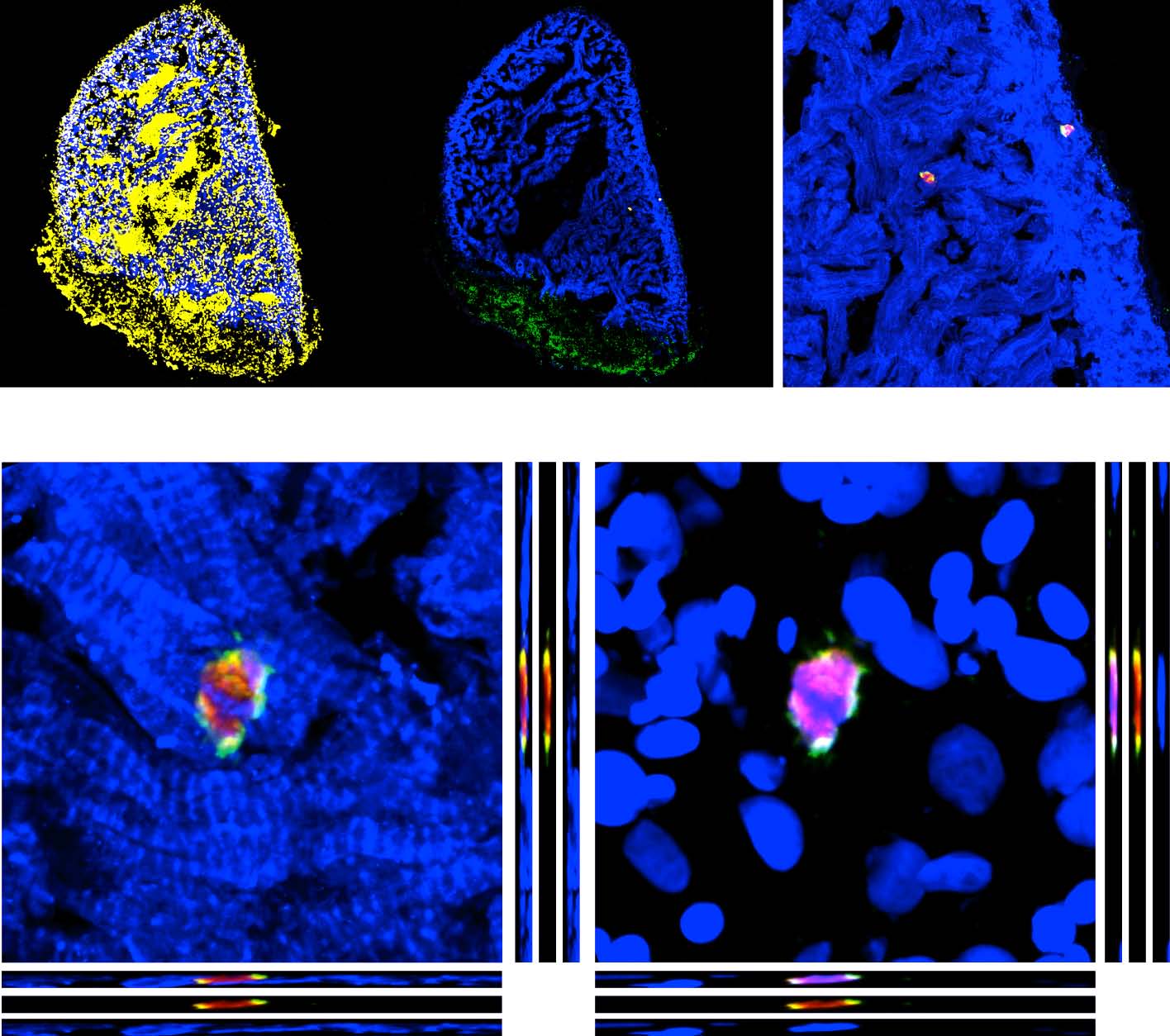
Mitotic CMs display upregulation of γ-tubulin.
(A-B) Representative image of a heart from cmlc2::EGFP transgenic fish at 14 dpci. Post-infarct tissue is encircled with a dashed line.
(B′) Mitotic CMs were detected by the PH3 immunostaining (red). PH3-positive CMs enhance the expression of γ-tubulin (green), which is a component of the mitotic spindle apparatus.
(C) Orthogonal projections of a mitotic CM demonstrate a polarized distribution of γ-tubulin around the PH3-positive chromosomes. Scale bar (A, B′, C) = 50 µm.The low level of γ-tubulin in centrosomes of non-mitotic cells is not visible with these image acquisition settings because the levels were optimized according to the fluorescence intensity of PH3-positive cells.
Reprinted from Developmental Biology, 399(1), Sallin, P., de Preux Charles, A., Duruz, V., Pfefferli, C., Jazwinska, A., A dual epimorphic and compensatory mode of heart regeneration in zebrafish, 27-40, Copyright (2015) with permission from Elsevier. Full text @ Dev. Biol.