
|
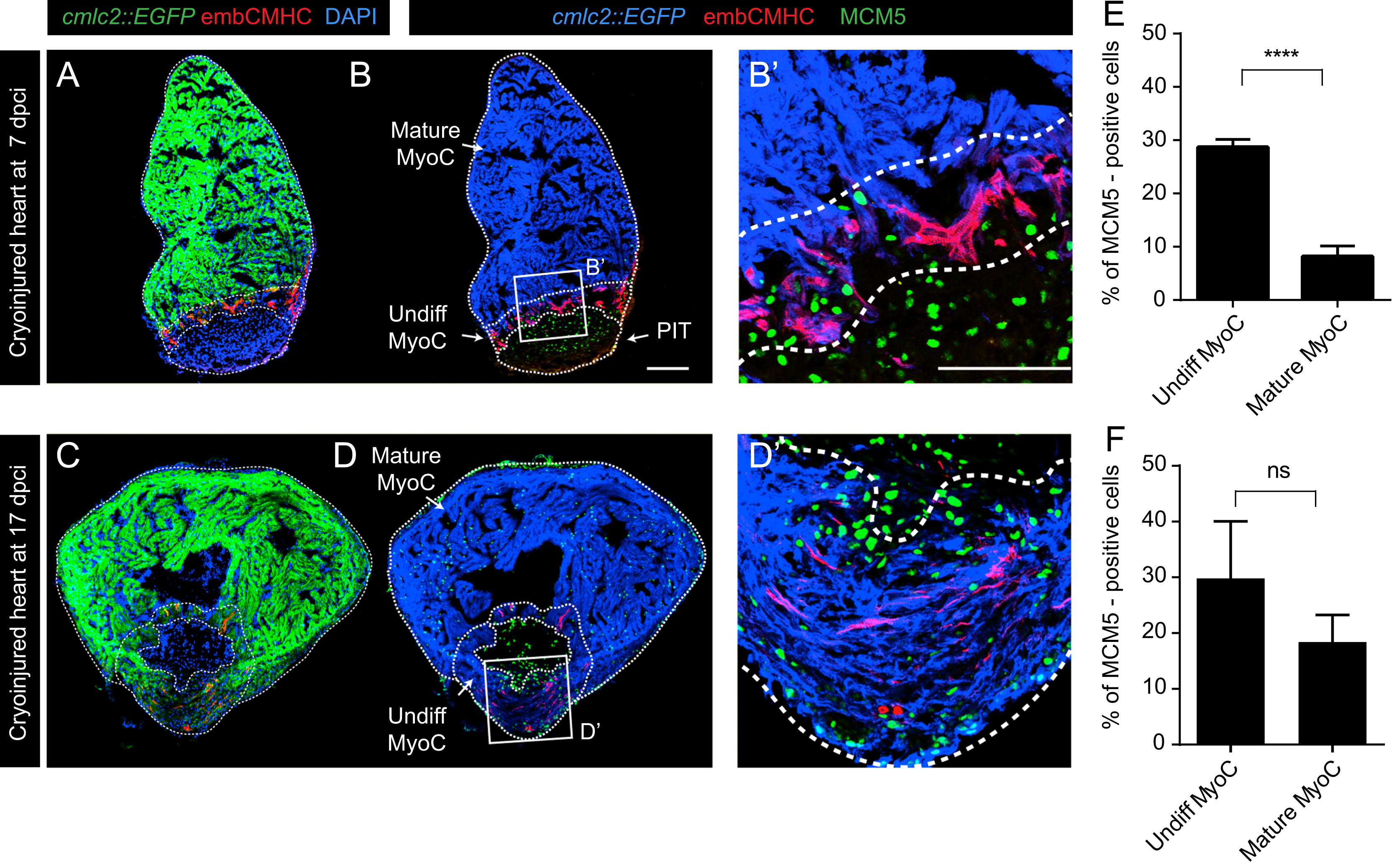
Fig. 7
Undifferentiated CMs display a higher proliferation rate than the mature CMs of the regenerating myocardium. (A–D) Representative images of cmlc2::EGFP hearts at 7 (A, B) and 17 (C, D) dpci. Post-infarcted tissue (PIT), undifferentiated myocardium (undiff. MyoC, embCMHC-positive, red) and mature myocardium (mature MyoC, embCMHC-negative) are encircled with a dashed line. Proliferating MCM5-positive cells were normalized to DAPI in each of the compartments. (B′) Higher magnification of the framed image in (B). A large accumulation of proliferating cells was observed in the undifferentiated myocardium compartment in comparison to the mature myocardium at 7 dpci. Scale bar (B, B100=′ µm. (E, F) Quantification of MCM5-positive cells reveals a significant increase in the proportion of proliferating cells in the undifferentiated myocardium as compared to mature myocardium at 7 dpci. At 17 dpci, the number of MCM5-positive cells is similar in both myocardial compartments. All results are expressed as the mean±standard error of the mean (S.E.M.) (n≥3 hearts, **** P-value<0.0001).
Reprinted from Developmental Biology, 399(1), Sallin, P., de Preux Charles, A., Duruz, V., Pfefferli, C., Jazwinska, A., A dual epimorphic and compensatory mode of heart regeneration in zebrafish, 27-40, Copyright (2015) with permission from Elsevier. Full text @ Dev. Biol.