
|
Fig. 4
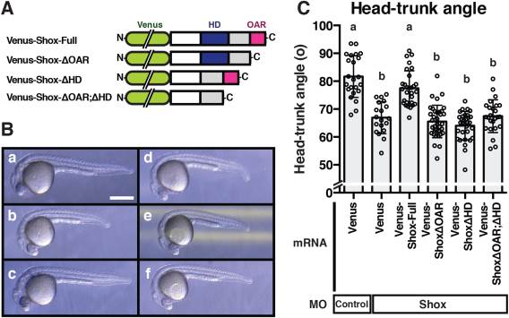
The in vivo functional characterization of Shox. A: Illustrations of varieties of deletion mutants of Shox. The synthesized N-term Venus fusion Shox mRNAs were all Shox MO resistant. The capped mRNAs (750 pg/embryo) were co-injected with Shox MO (8 ng/embryo). B: Representative embryos are displayed in a–f. Bar = 0.5 mm. a: Control MO and Venus mRNA; b: Shox MO and Venus mRNA; c: Shox MO and Venus-Shox-full mRNA; d: Shox MO and Venus-ShoxΔOAR mRNA; e: Shox MO and Venus-ShoxΔHD; f: Shox MO and Venus-ShoxΔOAR;ΔHD. C: The head-trunk angle of injected embryos at 26hpf stage. Values are represented as mean±S.D. (n=19–36). Groups with different letters are significantly different from each other (P< 0.05).