Fig. 2
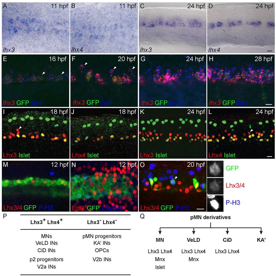
lhx4 and lhx3 are expressed in many cells in the ventral neural tube. All images oriented with anterior to the left. (A,B) Dorsal view of 11hpf embryos. Both lhx3 (A) and lhx4 (B) are expressed beginning at 11hpf in two medial cell stripes. (C,D) Lateral view of spinal cord adjacent to somites 8-12 of 24hpf embryo. Both lhx3 (C) and lhx4 (D) are expressed in the pMN domain; however, lhx4 is expressed in fewer cells than lhx3. (E-H) lhx3 and lhx4 expression in ventral neural tube of Tg(nrp1a:GFP) embryos. (E) At 16hpf, lhx3 and lhx4 are expressed at the dorsoventral level of GFP+ MNs, whereas some GFP+ cells express only lhx3 (arrowheads). (F) At 20hpf, lhx3 and lhx4 expression has expanded to include more dorsal GFP cells (arrowheads). (G,H) At 24hpf and 28hpf, lhx3 and lhx4 are broadly co-expressed in GFP+ and GFP cells throughout the lateral half of ventral spinal cord. (I-L) Lateral views of spinal cord hemisegments adjacent to somites 8-12 co-labeled for Islet and either Lhx3 or Lhx4. Dorsal Islet+ cells are Rohon–Beard sensory neurons. Anti-Islet labels PMNs at 18hpf, and PMNs plus a few SMNs at 24hpf. (I) At 18hpf, anti-Lhx3 labels all Islet+ PMNs as well as slightly more dorsal cells (arrowhead) in ventral neural tube. (J) At 18hpf, anti-Lhx4 labels almost exclusively Islet+ PMNs. (K) By 24hpf, anti-Lhx3 labels all Islet+ PMNs and SMNs and an increased number of cells in ventral neural tube. (L) Anti-Lhx4 also labels all Islet+ PMNs and SMNs in addition to other more dorsal cells (arrowhead) in ventral neural tube at 24hpf. (M) Lateral view of 12hpf Tg(olig2:GFP) embryo co-labeled with anti-Lhx3/4 and anti-phospho-Histone H3 (P-H3). (N) Dorsal view of 12hpf Tg(olig2:GFP) embryo incubated with EdU from 11hpf and labeled with EdU and anti-Lhx3/4. (O) Lateral view of 20hpf Tg(vsx1:GFP) embryo co-labeled with anti-Lhx3/4 and anti-P-H3. Enlarged, single channel images of indicated cell (arrowhead) demonstrate co-expression. (P) Summary of Lhx3- and Lhx4-expressing cells in the ventral spinal cord. (Q) Combinatorial expression of three transcription factors that uniquely label all four neuronal derivatives of the zebrafish pMN domain. Scale bars: 20µm.

