Fig. 2
Cell Proliferation and Survival in the Axial Vessels Do Not Appear to Be Affected by Gain or Loss of Function of Ifng1-2 Signaling
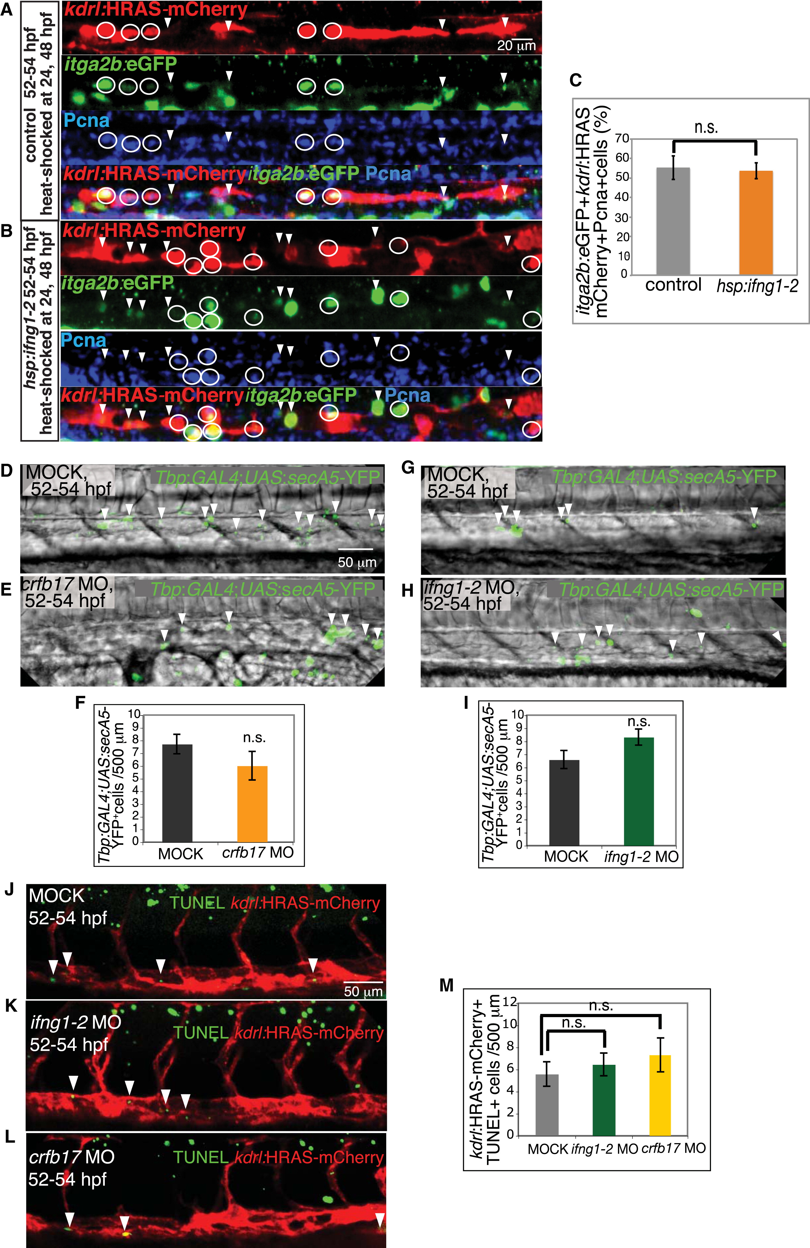
(A–C) ifng1-2 overexpression increases HSC number without affecting cell division. HSCs (itga2b:EGFP+kdrl:HRAS-mCherry+, green+red+) that exit the G0 phase, labeled by PCNA immunostaining (blue), in control embryos not harboring the Tg(hsp:ifng1-2-V5) transgene (A), and Tg(hsp:ifng1-2-V5) embryos (B) are indicated by white circles. PCNA HSCs are marked with white arrowheads. (C) Percentage of PCNA+HSCs per total HSCs in the dorsal aorta. Values represent means ± SEM, n = 9 embryos per group, n.s., not significant (p > 0.05).
(D–M) Ifng1-2 and Crfb17 knockdown has no effect on apoptosis. Apoptotic cells (green, white arrowheads) visualized by Tg(Tbp:GAL4;UAS:secA5-YFP) expression (D–I) and TUNEL assay (J–M) in the axial vessels of MOCK-injected (D, G, and J), crfb17 MO-injected (E and L), and Ifng1-2 MO-injected (H and K) embryos at 52–54 hpf. Numbers of Tbp:GAL4;UAS:secA5-YFP+ cells (F and I) and TUNEL+kdrl:HRAS-mCherry+ cells per 500 µm dorsal aorta length (M) are shown as means ± SEM, n = 11–25 embryos, n.s. p > 0.05. All images are lateral views, dorsal up and anterior to the left.
Reprinted from Developmental Cell, 31, Sawamiphak, S., Kontarakis, Z., Stainier, D.Y., Interferon Gamma Signaling Positively Regulates Hematopoietic Stem Cell Emergence, 640-653, Copyright (2014) with permission from Elsevier. Full text @ Dev. Cell

