Fig. 1
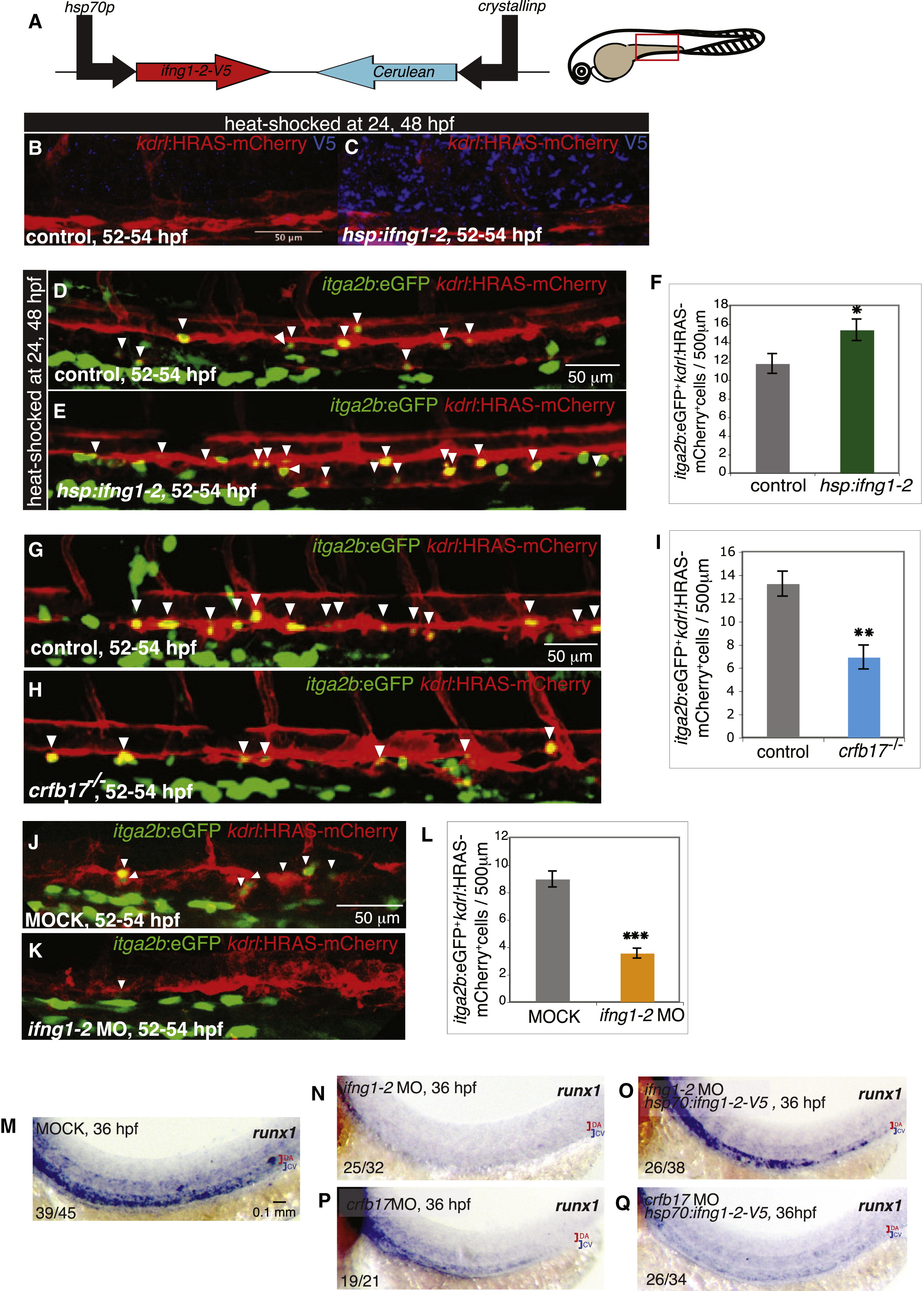
Ifng1-2 and Its Receptor Crfb17 Positively Regulate HSC Development
(A) Schematic drawing of construct for heat-shock-inducible expression of ifng1-2. The area imaged and analyzed in all experiments (red box) is shown in the embryo illustration.
(B and C) Immunofluorescent labeling of V5-tagged Ifng1-2 (blue) in the vicinity of the AGM of control animals lacking the hsp:ifng1-2-V5 transgene (B) compared with Tg(hsp:ifng1-2) animals (C). The AGM region is recognized by Tg(kdrl:HRAS-mCherry) expression (red) in the vasculature.
(D and E) Increased number of HSCs at the ventral wall of the dorsal aorta and in the cardinal vein lumen upon ifng1-2 overexpression. HSCs (white arrowheads) in control (D) and Tg(hsp:ifng1-2-V5) (E) embryos were fluorescently labeled by Tg(itga2b:EGFP) (green) and Tg(kdrl:HRAS-mCherry) (red) expression.
(F) Number of HSCs per 500 µm aortic length. Values represent means ± SEM, n = 21–22 embryos. p d 0.05. All embryos were heat-shocked at 24 and 48 hpf and imaged at 52–54 hpf. Of note, the numbers of Tg(itga2b:EGFP)+;Tg(kdrl:HRAS-mCherry) (only green) pronephric duct cells appear unaffected by Ifng1-2 overexpression.
(G–I) Impaired HSC development in crfb17-/- embryos. Control (crfb17+/+) (G) and crfb17-/- (H) siblings were imaged at 52–54 hpf, and itga2b:EGFP+kdrl:HRAS-mCherry+ HSC (white arrowheads) numbers were analyzed prior to genotyping. (I) HSCs per 500 µm aortic length. Values represent means ± SEM, n = 8–14 embryos, p d 0.01.
(J–L) Ifng1-2 knockdown recapitulates the HSC phenotype of crfb17 mutants. itga2b:EGFP+kdrl:HRAS-mCherry+ HSC (white arrowheads) in mock (1% phenol red in nuclease-free distilled water)-injected (J) or ifng1-2 MO-injected (K) embryos imaged at 52–54 hpf. Numbers of HSCs per 500 µm aortic length is shown in (L). Values represent means ± SEM, n = 32–40 embryos, p d 0.001.
(M–O) Reduction of runx1 expression upon Ifng1-2 knockdown is restored by ifng1-2 induction. Expression of the HSC marker runx1 in mock-injected (M) and ifng1-2 MO-injected embryos without (N) and with (O) the hsp70:ifng1-2-V5 transgene.
(P and Q) ifng1-2 overexpresssion is unable to rescue Crfb17 knockdown. runx1 expression in crfb17 MO-injected embryos without (P) and with (Q) the hsp70:ifng1-2-V5 transgene. All embryos were heat shocked at 24 hpf, and runx1 expression was assessed by in situ hybridization at 36 hpf. The number of embryos showing the representative phenotype per total number of embryos analyzed is indicated in the lower left corner. Red brackets identify the dorsal aorta (DA); blue brackets identify the cardinal vein (CV). All images are lateral views, dorsal up, anterior to the left.
Reprinted from Developmental Cell, 31, Sawamiphak, S., Kontarakis, Z., Stainier, D.Y., Interferon Gamma Signaling Positively Regulates Hematopoietic Stem Cell Emergence, 640-653, Copyright (2014) with permission from Elsevier. Full text @ Dev. Cell

