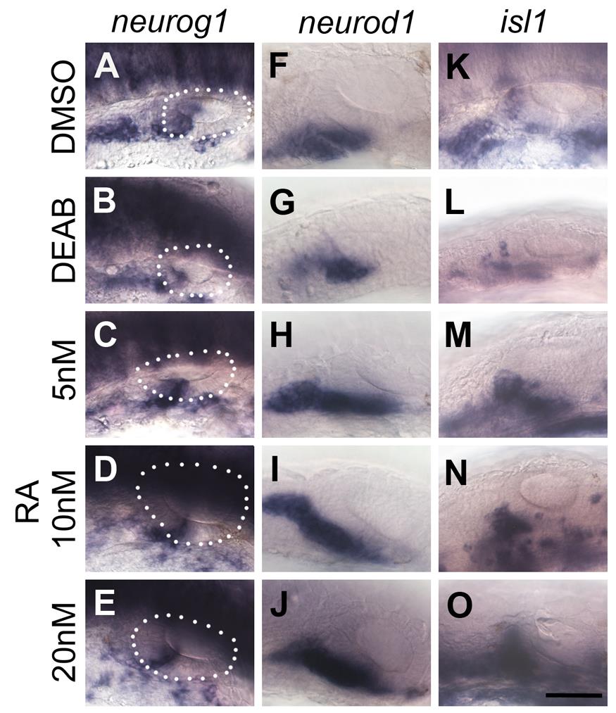
Fig. S8 A role for RA in regulating zebrafish otic neurogenesis. Embryos were treated with DMSO, DEAB or RA from 18/20S to 26 hpf. (A–E) The dotted line demarcates the OV. Wild-type embryos treated with DMSO show normal expression of neurog1 (A), while expression is decreased in the OV of wild-type embryos treated with DEAB (B). Otic expression of neurog1 is relatively normal, or slightly reduced, in embryos treated with 5 nM (C), 10 nM RA (D) and 15 nM (E) RA. (F–J) Embryos treated with DMSO show normal expression of neurod1 (F), while expression is decreased in the OV of embryos treated with DEAB (G), and increased in embryos treated with 5 nM (H), 10 nM (I) and 15 nM (J) RA. (K–O) Embryos treated with DMSO show normal otic expression of isl1 (K), while expression is decreased in embryos treated with DEAB (L), and increased in embryos treated with 5 nM (M), 10 nM (N) and 15 nM (O) RA. All panels are lateral views with anterior to the left. Scale bar: 50 µm.
Image
Figure Caption
Acknowledgments
This image is the copyrighted work of the attributed author or publisher, and
ZFIN has permission only to display this image to its users.
Additional permissions should be obtained from the applicable author or publisher of the image.
Full text @ PLoS Genet.

