
|
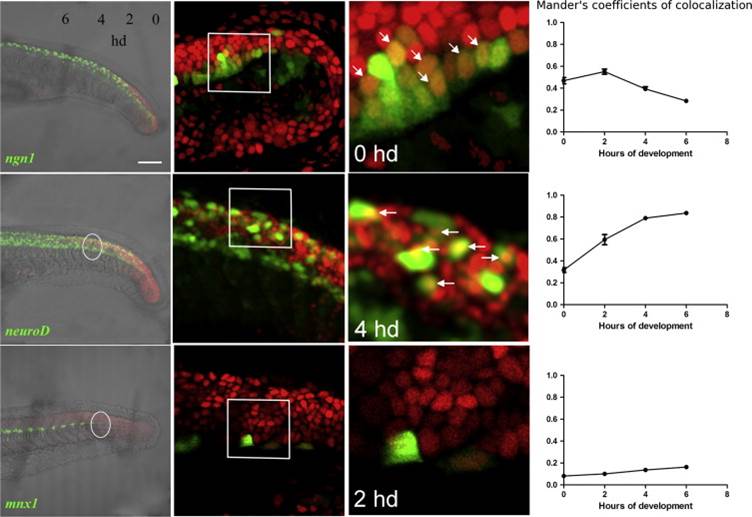
Fig. 5 Smad3 reporter is expressed in progenitors and precursors. Confocal lateral view of tail of double transgenic embryos obtained crossing Tg(12xSBE:nls-mCherry)ia15 to the following transgenics: Tg(ngn1:GFP)sb1, Tg(2.4 kb neurod:EGFP) and Tg(mnx1:GFP)ml2. Colocalization was measured in the tail of 24 hpf double transgenic embryos with the following method. From 15 to 24 hpf, two somites are formed each hour. Tail region was divided in pairs of somites and colocalization (Manders′ coefficient referred to TGFβ-mCherry fluorescence) evaluated in four of them (as white dotted circles) starting from the edge of the tail toward the trunk. For each time point, the average value of Manders′ coefficient has been calculated from six double transgenic embryos and plotted on as a function of the corresponding hour of development (hd). Second column: magnification of specific regions (single plane). Third column: double fluorescent cells are shown with arrowheads (single plane). Developmental time of each magnification is indicated inside the panel. Fourth column: graphical representation of quantitative analysis as Manderós coefficients of the four developmental points; n=6 per each point. Scale bar is 100 µm in the first column, 20 µm in the second column, 10 µm in the last column.
Reprinted from Developmental Biology, 396(1), Casari, A., Schiavone, M., Facchinello, N., Vettori, A., Meyer, D., Tiso, N., Moro, E., Argenton, F., A Smad3 transgenic reporter reveals TGF-beta control of zebrafish spinal cord development, 81-93, Copyright (2014) with permission from Elsevier. Full text @ Dev. Biol.