
Image
|
Figure Caption
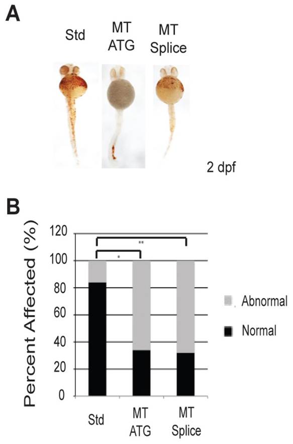
Fig. 3
Loss of etv7 causes a marked reduction in hemoglobinized red blood cells. (A) etv7 morphants stained with o-dianisidine at 2 dpf. Std represents embryos injected with 8.2 ng of standard control morpholino, MT ATG are embryos injected with 8.2 ng of translation-blocking morpholino, and MT Splice are embryos injected with 4.1 ng of splice-site morpholino. Normal embryos were classified by the presence of adequate heme staining according to o-dianisidine, and fish with reduced heme staining are classified as abnormal. (B) Quantitation of A (MT ATG n=44, Std n=44, MT Splice n=34). *Pd0.0001, **Pd0.0001.
Figure Data
Acknowledgments
This image is the copyrighted work of the attributed author or publisher, and
ZFIN has permission only to display this image to its users.
Additional permissions should be obtained from the applicable author or publisher of the image.
Full text @ Dis. Model. Mech.