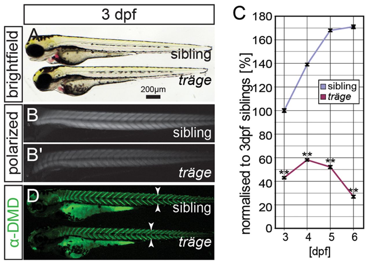
Fig. 1 The mutant träge (trg) shows a reduction in birefringence, indicating muscle damage. (A) Under brightfield microscopy, trg mutants appear similar to their wild-type siblings. (B) Under polarised light, the muscle of siblings appears brighter than that of the (B′) trg mutants due to a reduction in birefringence. (C) Quantification of the birefringence followed by normalization to that of 3-dpf-old siblings reveals that the birefringence of the siblings increases from 3 dpf to 6 dpf, roughly following a sigmoidal curve. By contrast, 3-dpf- to 6-dpf-old trg larvae show a highly significant reduction in birefringence when compared with that of 3-dpf siblings (P<0.01, n=3). (D) Immunohistochemistry with antibodies against dystrophin shows that dystrophin expression at the vertical myosepta (arrowheads) is unaffected in trg mutants. Data are means ± s.e.m., **P<0.01. Scale bar: 200 μm.
Image
Figure Caption
Figure Data
Acknowledgments
This image is the copyrighted work of the attributed author or publisher, and
ZFIN has permission only to display this image to its users.
Additional permissions should be obtained from the applicable author or publisher of the image.
Full text @ Dis. Model. Mech.

