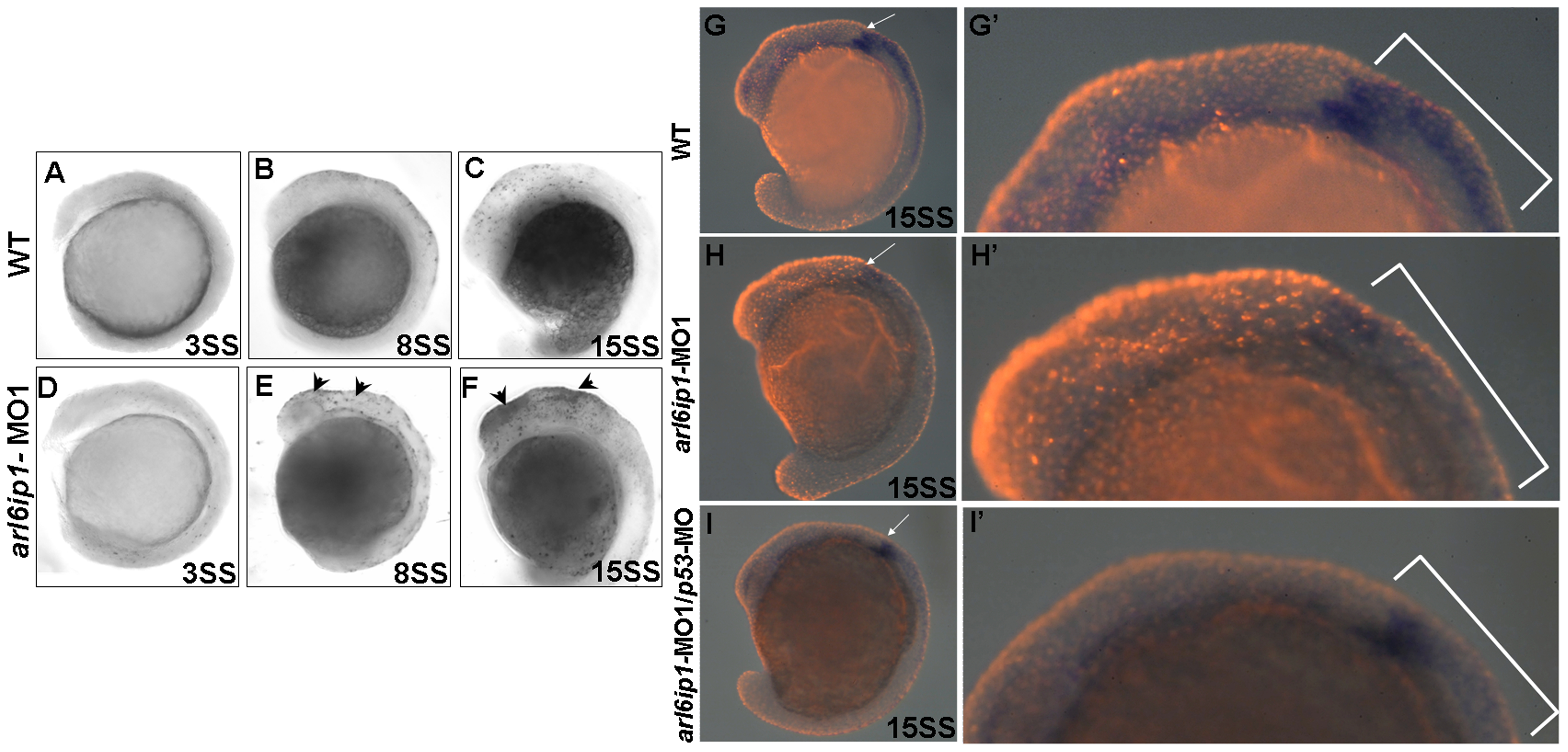
Fig. 8 Cell death does not cause the loss of neural crest cells.
Lateral views of TUNEL-labeled wild-type (WT) embryos (A-C) or arl6ip1-MO1-injected embryos (D-F) at 3-somite stage (A, D), 8-somite stage (B, E), and 15-somite stage (C, F), respectively. (A, D) There were no TUNEL-positive cells in WT and arl6ip1-MO1 embryos. (B, C, E, F) Compared to WT embryos, arl6ip-deficient embryos displayed a significant increase of TUNEL-positive cells in the dorsal region of embryos at the 8- and 15-somite stages (indicated by arrowheads) (B and C vs. E and F). (G, H, I) Double-labeling analysis with crestin (dark blue) and TUNEL (red fluorescent) showed that only limited cell death occurred at the expression region of crestin. (G′, H′, I′) The views of the embryos shown in panels G′, H′ and I′ were at higher magnification.

