
|
Fig. S6
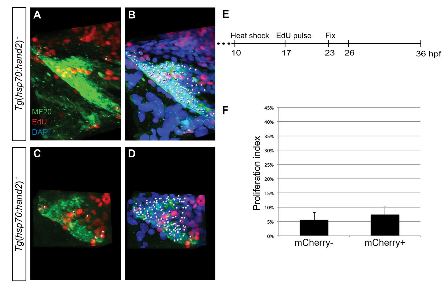
No evident influence of hand2 overexpression on cardiomyocyte proliferation within the heart tube at 23 hpf. (A-D) EdU incorporation in hearts of (A,B) nontransgenic and (C,D) Tg(hsp70:hand2) embryos at 23 hpf, following heat shock at 10 hpf and EdU pulse at 17 hpf; partial reconstructions of confocal z-stacks. Images depict the elongating cardiac cone, positioned with its arterial end toward the top. (A,C) White dots indicate EdU-positive (red) cells that are also MF20-positive (green) differentiated cardiomyocytes. (B,D) White dots indicate all nuclei (DAPI, blue) of myocardial cells, including both EdU-positive and EdU-negative cardiomyocytes. (E) Timeline of experimental design. (F) Bar graph compares proliferation indexes in nontransgenic (mCherry- negative) and Tg(hsp70:hand2) (mCherry-positive) embryos, as in Fig. 5H. No change in proliferation index is seen in hand2- overexpressing cardiomyocytes (n=10-11; p=0.252). Similarly, when we assessed EdU incorporation in hand2-overexpressing embryos at 26 hpf, following heat shock at 10 hpf and EdU pulse at 14 hpf, we did not see an increased proliferation index in hand2- overexpressing cardiomyocytes (proliferation index of 28 ± 4% in nontransgenic embryos compared to proliferation index of 27 ± 4% in hand2-overexpressing embryos; n=7-10, p=0.58).