
|
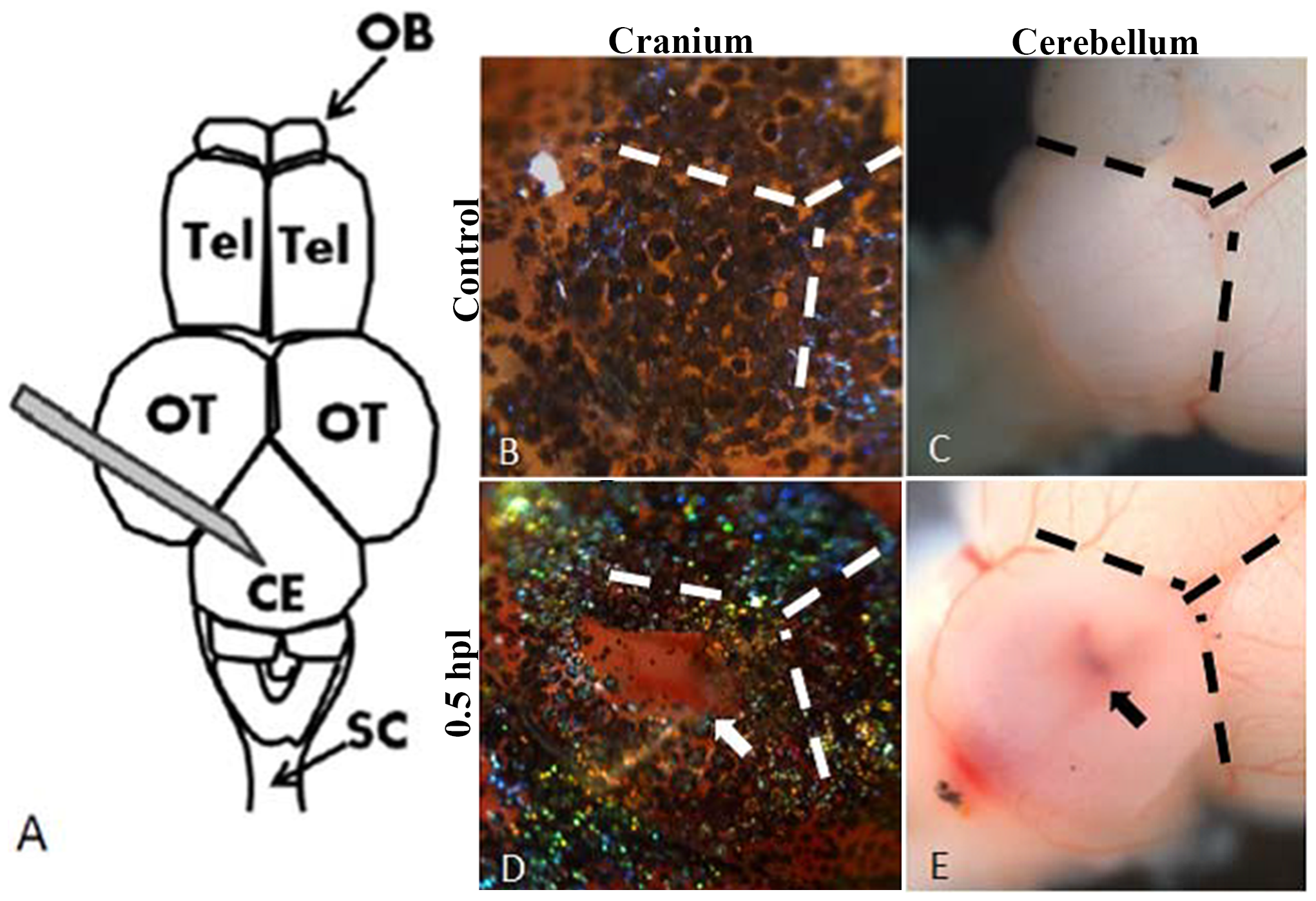
Fig. 1
The diagram of the stab lesion assay.
A: Schematic diagram of the stab lesion. A 27G syringe was used to create a cranial wound of depth 1.5 mm in the cerebellum (OB, olfactory bulb; Tel, telencephalon; OT, optic tectum; Ce, cerebellum; SC, spinal cord). B–E: Bright field images of the cranium (B, D) and exposed brains (C, E) of control fish and after the stab lesion. B and C show intact cranium and cerebellum of the control (no stab lesion) and D and E show the injured cranium and cerebellum of an experimental animal at 0.5 hour post-lesion (hpl). The fresh wound can be seen clearly on both the cranium and the cerebellum (D, E; the white arrow shows the lesion site on the cranium and the black arrow shows the wound on the exposed cerebellum).