
|
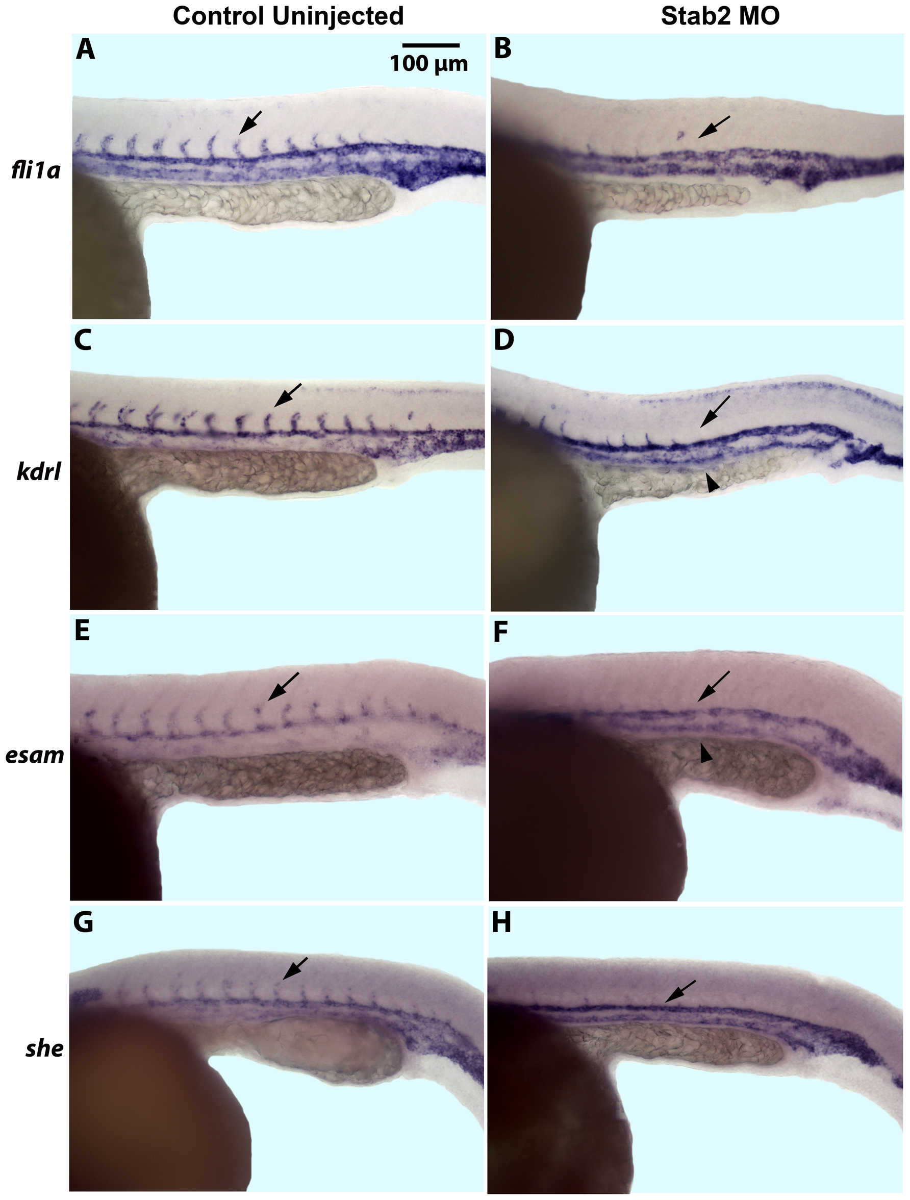
Fig. 1
Stab2 morphants display a lack of intersegmental vessels.
(A–H) In situ hybridization analysis shows a lack of ISVs in Stab2 morphants as observed by endothelial marker fli1a (A and B), kdrl (C and D), esam (E and F), and she (G and H) expression at 24 hpf, as compared to uninjected controls. Arrows indicate ISVs in control embryos and missing ISVs in morphants. Arrowheads indicate stronger PCV expression of kdrl and esam in morphant embryos (D and F). Lateral view, anterior is to the left; trunk and tail region is shown. Morphants were injected with a cocktail containing Stab2 MO1 and MO2, as well as p53 MO. ISVs: intersegmental vessels; PCV: posterior cardinal vein.