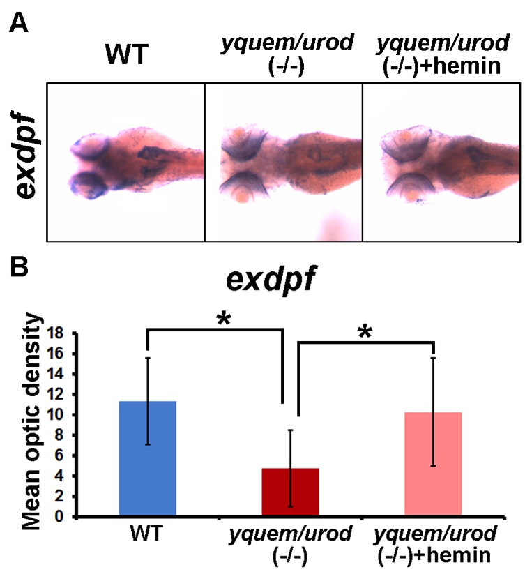
Fig. S7 (A) Representative images of in situ hybridization staining indicate 127 downregulation of exdpf in zebrafish yquem/urod (-/-). Despite the expression of 128 exdpf elsewhere in larvae, it is the exocrine pancreatic expression that is most affected 129 in the mutant fish. Dorsal view, anterior to left. (B) Mean optic densities of in situ 130 hybridization staining of a group of larvae (10-12 each) corresponding to Fig. S7A 131 quantified with ImageJ. Student’s t tests were conducted. * P<0.05. These in situ 132 hybridization results are consistent with the qRT-PCR results shown in Fig. 7B. All 133 larvae were at 84 HPF (hours postfertilization).
Image
Figure Caption
Figure Data
Acknowledgments
This image is the copyrighted work of the attributed author or publisher, and
ZFIN has permission only to display this image to its users.
Additional permissions should be obtained from the applicable author or publisher of the image.
Full text @ Dis. Model. Mech.

