
Image
|
Figure Caption
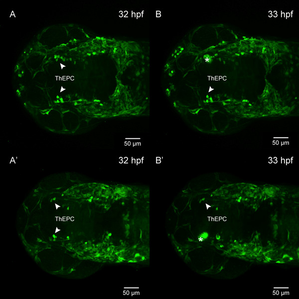
Fig. S2
Ablation of ThEPC cells at 32 hpf, related to Figure 2.
(a-b′) Dorsal view, anterior to the left, MIP of Et(-1.0otpa:mmGFP)hd1 transgenic embryos before and after ThEPC cell ablation. (a-a′) White arrowheads mark the ThEPCs before ablation. (b-b′) Asterisks mark the site of ablation, white arrowheads mark the non-ablated ThEPC.The original stacks were cropped and the gamma was corrected to 0.60 for display purposes.
ThEPC, thalamic-epithalamic early projecting cluster.
Acknowledgments
This image is the copyrighted work of the attributed author or publisher, and
ZFIN has permission only to display this image to its users.
Additional permissions should be obtained from the applicable author or publisher of the image.
Full text @ Neural Dev.