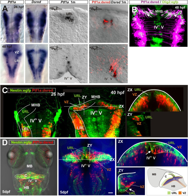
Fig. 2 Distinct cerebellar progenitor populations are established early in the embryo. (A) Expression of ptf1a, DsRed (mRNA) and DsRed (protein) in developing and juvenile Ptf1a:DsRed transgenic fish. Ptf1a, DsRed and DsRed show similar expression patterns; (B) In vivo expression of DsRed and Egfp in the embryonic and juvenile Ptf1a:DsRed and Olig2:egfp transgenic fish. Overlapping Egfp and DsRed expression is seen in the VZ of the cerebellar primordium; (C) Nestin:egfp+ (green) and Ptf1a:DsRed+ (red) progenitors in the cerebellar primordium. The Nestin:egfp labels cells in the URL, while Ptf1a:DsRed line labels cells in the VZ. Two days post-fertilization Nestin:egfp+ and Ptf1a:DsRed+ cells form distinct populations in the URL and VZ of the cerebellar primordium; (D) A dorsal overview of Nestin:egfp+ (green) and Ptf1a:DsRed+ (red) progenitors in the hindbrain of a 5-day-old larval zebrafish. Two distinct progenitor domains are visible in the cerebellum (the junction is indicated with an arrow). Ptf1a:DsRed+ cells localize along the ventricular zone of the IVth ventricle (the VZ domain is labeled with a hatched orange line), while Nestin:egfp+ cells localize in the URL (hatched green line). MHB: Mid-hindbrain boundary; LRL: Lower rhombic lip.
Image
Figure Caption
Figure Data
Acknowledgments
This image is the copyrighted work of the attributed author or publisher, and
ZFIN has permission only to display this image to its users.
Additional permissions should be obtained from the applicable author or publisher of the image.
Full text @ Neural Dev.

