
|
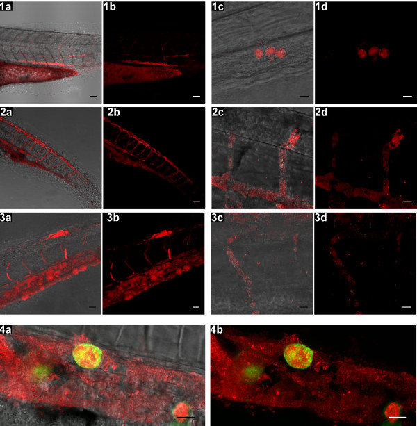
Fig. 2
Invasion of S. epidermidis into the zebrafish embryo body. Confocal z-stacks are shown as transmission/fluorescence overlay (a &c) and fluorescence images (b &d). Panel 1: at 3 DPI mCherry labelled S. epidermidis is observed inside the body (1a &1b, scale bar: 50 µm), and intracellular in the hematopoietic region (1c &1d, scale bar: 10 µm). Panel 2: at 4 DPI bacteria are found inside the vasculature (2a &2b, scale bar: 50 µm), including the intersegmental vessels (2d &2d, scale bar: 10 µm). Panel 3: at 5 DPI bacteria are still persisting in the vasculature (3a &3b, scale bar: 25 µm) and in the intersegmental vessels (3c &3d, scale bar: 10 µm). Panel 4: bacteria being taken up by mpeg1:KAEDE positive cells and extracellular in the hematopoietic region at 3 DPI (scale bar: 10 µm).