
|
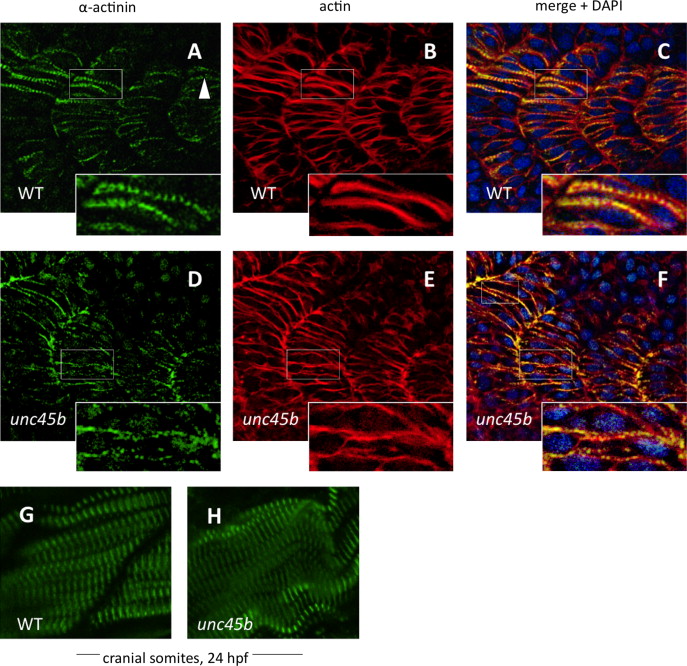
Fig. 7
Nucleation of α-actinin is delayed in zebrafish unc45b mutants. Immunofluorescent staining of WT (A–C) and unc45b mutant embryos (D–F) at 20 h post-fertilization, displaying the 19th to 22nd somites. The caudal-most somites of these embryos were still undergoing myogenesis, as shown by the incomplete elongation of myofibers and localization of α-actinin and actin staining in WT embryos (A and B, inserts). Nucleation of α-actinin is the first indication of periodic myofibril patterning (arrowhead). In mutant embryos, organization of α-actinin at costamere attachment sites was not yet complete, and nucleation had just begun (D, insert). Actin counter-staining with phalloidin (B, E) demonstrates the ongoing organization of actin in early myofibers, which can be compared with the pattern of α-actinin localization in merged images (C, F). Blue fluorescence indicates DAPI nuclear stain. The cranial-most somites at 24 hpf display relatively normal patterns of α-actinin staining in mutants (H) compared to WT embryos (G).
Reprinted from Developmental Biology, 390, Myhre, J.L., Hills, J.A., Jean, F., Pilgrim, D.B., Unc45b is essential for early myofibrillogenesis and costamere formation in zebrafish, 26-40, Copyright (2014) with permission from Elsevier. Full text @ Dev. Biol.