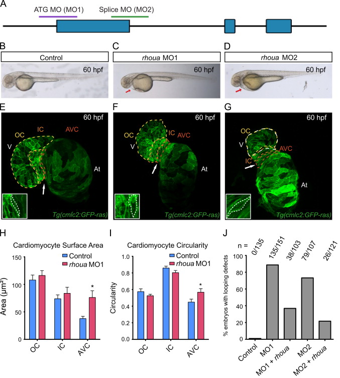
Fig. 2 Loss of rhoua results in cardiac edema due to morphologic atrioventricular (AV) canal defects. (A) The following regions of rhoua were targeted to create the rhoua ATG (MO1) and splice (MO2) morpholinos (MO). (B–D) Brightfield microscopy imaging shows that rhoua ATG and splice MO injected embryos (C, D) exhibited cardiac edema (red arrows) by 60 hpf, but control MO injected embryos (B) did not. (E–G) Confocal projections of 60 hpf Tg(cmlc2:ras-eGFP) (E) control MO, (F) rhoua ATG MO1, and (G) rhoua splice MO2 injected zebrafish revealed that rhoua MO injected zebrafish hearts failed to undergo proper cardiac looping and displayed AV canal defects. White arrow points to AV canal. Outer curvature (OC), inner curvature (IC), and atrioventricular canal (AV) are outlined in yellow, orange, and red dashed lines, respectively. At– atrium, V – ventricle. Inset shows representative AV cardiomyocytes, which appear more rounded and larger in MO injected embryos compared to control embryos. (H, I) Bar graphs represent cardiomyocyte (H) surface area and (I) morphology/circularity measurements of control and ATG MO1 Tg(cmlc2:ras-eGFP) hearts at 60 hpf. Mean+s.e.m. Studentós t-test, *p<0.05 (n=5 control and 7 rhoua morpholino injected zebrafish hearts). (J) 88.7% and 73.3% of zebrafish embryos injected with a rhoua ATG (MO1) and splice (MO2) MOs exhibited cardiac edema and AV canal defects, but wild-type embryos injected with control MO did not (0%). Injection of 150 pg of wild-type zebrafish rhoua was able to rescue the cardiac phenotype in the rhoua ATG and splice MO injected zebrafish and reduce the mutant phenotype to 36.9% and 21.7% for ATG MO1 (MO1+rhoua) and splice MO2 (MO2+rhoua), respectively. n indicates the number of embryos with cardiac phenotype/total number of embryos injected for each condition.
Reprinted from Developmental Biology, 389, Dickover, M., Hegarty, J.M., Ly, K., Lopez, D., Yang, H., Zhang, R., Tedeschi, N., Hsiai, T.K., Chi, N.C., The atypical Rho GTPase, RhoU, regulates cell-adhesion molecules during cardiac morphogenesis, 182-91, Copyright (2014) with permission from Elsevier. Full text @ Dev. Biol.

