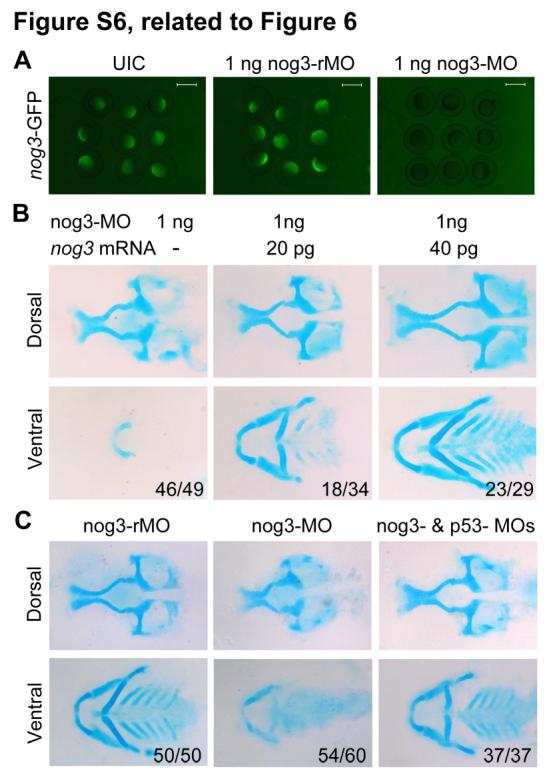
Fig. S6 nog3-MO-induced head skeletal defects could be rescued by co-injection of p53-MO, related to Figure 6. (A) Effectiveness of nog3-MO. Embryos injected with 50 pg nog3-GFP plasmid DNA showed green fluorescence at the shield stage. Green fluorescence was obviously decreased when co-injected with 1 ng nog3-MO. (B) co-injection of nog3 mRNA rescued nog3-MO-induced pharyngeal cartilage defects. Embryos at the one-cell stage were injected with 1 ng of nog3-MO alone or plus an indicated amount of nog3 mRNA and harvested at 80 hpf for staining head cartilages with Alcian blue. nog3 mRNA carried several mutated bases around the translation start site, which did not change the identities of the encoded amino acids, so that nog-MO would not bind to it. (C) The head cartilage defects in nog3 morphants could be rescued by co-injection of 4 ng p53-MO. Head cartilages stained with Alcian blue at 80 hpf. Upper panel, lateral views; lower panel, dorsal views.
Reprinted from Developmental Cell, 24(3), Ning, G., Liu, X., Dai, M., Meng, A., and Wang, Q., MicroRNA-92a upholds Bmp signaling by targeting noggin3 during pharyngeal cartilage formation, 283-295, Copyright (2013) with permission from Elsevier. Full text @ Dev. Cell

