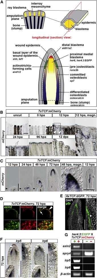
Fig. 1
Wnt/β-Catenin Pathway Activation during Zebrafish Caudal Fin Regeneration
(A) Cartoons summarizing relevant anatomical structures and expression domains of a fin regenerate during the outgrowth phase. Whole-mount and longitudinal section views are shown.
(B) mCherry RNA expression in 7xTCF:mCherry transgenic regenerates. Note transcript expression at 12 hpa in the interray tissue (arrowheads) and at 24 hpa distally to the bony ray (arrow).
(C) mCherry expression is confined to the mesenchyme at all stages analyzed. The basal epidermal layer is devoid of staining (asterisk).
(D) axin2 (green) and mCherry RNA (red) are coexpressed in the distal blastema (arrowhead) and interray cells (arrow) in 7xTCF:mCherry transgenic regenerates.
(E) gfp RNA expression in 6xTCF:dGFP transgenic regenerates is confined to the blastema. The basal epidermal layer is devoid of staining (asterisk).
(F) lrp5 transcripts are confined to proximal mediolateral domains of the blastema and are absent in the distal (arrowhead) blastema, the osteoblast progenitors (asterisk), and the basal epidermal layer (arrow). lrp6 transcripts are predominantly detected in the distal blastema (arrowhead). Asterisk: (pre)osteoblasts; arrow: basal epidermal layer.
(G) RT-PCR of indicated genes on cDNA derived from fluorescence-activated-cell-sorted 7xTCF:mCherry; her4.3:EGFP double transgenic regenerates at 72 hpa. Endogenous lrp5/6 transcripts are not detected in the fluorescence-negative, blastema-free cell fraction that contains epidermal cells whereas spry4 is.
In (B)–(G), confocal images of a whole-mount (D) or longitudinal section (E) are shown. Black arrowheads, amputation plane. Scale bars, 200 μm, 100 μm (D, whole mount),10 μm (D), and 100 μm (sections).

