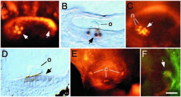
Fig. 2 Differentiation of hair cells in wild-type embryos. (A) Dorsolateral view of the otic vesicle at 30 h showing anti-Pax2 staining. The nuclei of presumptive hair cells stain intensely (arrows) whereas surrounding cells show lower staining levels. (B) Parasagittal section of a 30 h embryo stained in whole mount with anti-Pax2 and anti-acetylated tubulin antibodies. Hair cells in the utricular macula are marked by their strong nuclear Pax2 staining (arrow), as well as their stained kinocilia and associated otolith (o). Support cells show no detectable staining. (C) Dorsal view of the otic vesicle at 48 h showing anti-Pax2 staining in the utricular macula. The nuclei of hair cells stain intensely (arrow) and presumptive nascent hair cells (n) surrounding the macula show lower staining levels. (D) Longitudinal section of a 60 h embryo stained in whole mount with anti-Pax2 and anti-acetylated tubulin antibodies. In this slightly oblique dorsal view of the saccular macula, the kinocilia are not visible, but hair cells are clearly marked by their nuclear anti-Pax2 staining (arrow) and the close proximity of the otolith (o). Support cells are not labeled. (E) Lateral view of the otic vesicle at 60 h showing anti-Pax2 staining in the cristae (c). (F) Lateral view of posterior crista in a 60 h embryo stained with anti-Pax2 (red) and anti-acetylated tubulin (green). Hair cells (arrow) show nuclear Pax2 staining and ciliary acetylated tubulin staining. Support cells are not labeled. In all panels, anterior is to the right. (A,B,E,F) Dorsal is up; (C) medial is upward, and (D) medial is downward. Scale bar, 15 μm (F), 20 μm (A-D), or 65 μm (E).
Image
Figure Caption
Acknowledgments
This image is the copyrighted work of the attributed author or publisher, and
ZFIN has permission only to display this image to its users.
Additional permissions should be obtained from the applicable author or publisher of the image.
Full text @ Development

