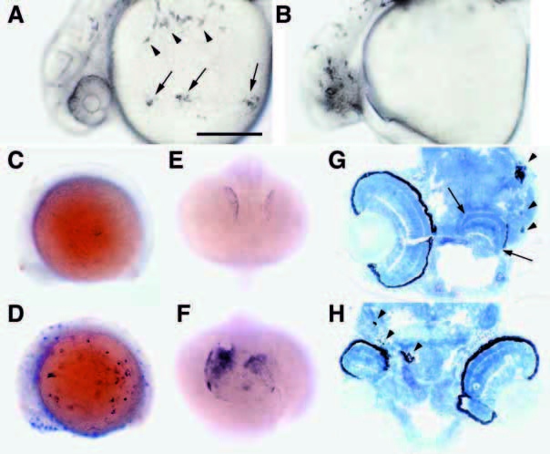
Fig. 9 Misexpression of nacre induces ectopic melanophores and disrupts eye development. (A,B) Wild-type embryos injected with nacre RNA at the 1- to 4-cell stage are shown at approximately 30 hpf. Embryos were frequently observed that displayed large patches of melanophores with abnormal morphology, as well as patches of similar cells in uncharacteristic locations such as the ventral yolk ball (A, arrows). Arrowheads indicate normal melanophores. Disorganization of one or both eyes in otherwise normal embryos was frequently observed (B). (C-F) Ectopic expression of nacre induces ectopic expression of trp2. Embryos were injected with pHSMT3A. 1-I219F (C) or pHS-MT3A.1 (D) and heat shocked from 10-12 hpf, then fixed at 14 hpf and processed for in situ hybridization with probes for melanophore marker trp2 (blue) and the neural crest marker fkd6 (orange). Widespread trp2-positive cells are observed in (D) prior to neural crest emigration. Injection of wild-type (F) but not mutant (E) nacre RNA expands the domain of trp2 expression in the optic primordium of some embryos at 18 hpf. (G,H) Examples of disrupted eye morphologies in frontal sections of 72 hpf embryos. The embryo in G has one approximately normal eye on the left, and a second laminated structure with a small amount of RPE on the right (arrows). The eyes in the embryo in H are of reduced size and show disorganization and irregularities in the RPE. Arrowheads, ectopic pigment cells. Scale bar, A,B, 250 μm; C-F, 300 μm; G,H, 100 μm.
Image
Figure Caption
Acknowledgments
This image is the copyrighted work of the attributed author or publisher, and
ZFIN has permission only to display this image to its users.
Additional permissions should be obtained from the applicable author or publisher of the image.
Full text @ Development

