
|
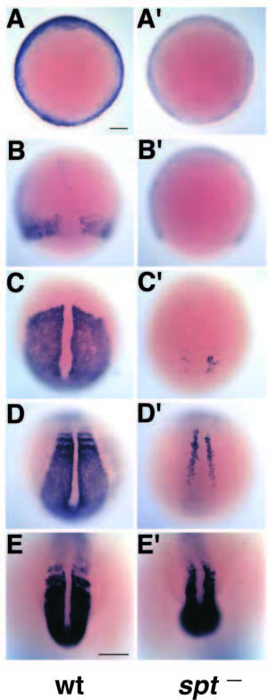
Fig. 3 papc expression is defective in spt mutant embryos. (A-E) Wild-type embryos. (A′-E′) sptb104 homozygous mutant embryos. (A,A′) Shield stage showing greatly reduced papc in spt- embryos at the onset of gastrulation. (B,B′) 80% epiboly showing lack of papc expression in the marginal zone of the spt mutant. (C,C′) Bud (10 hour) stage, (D,D′) 4-6 somite stage; expression is detected only in a few adaxial-like cells in spt mutants. (E,E′) 18 somite stage; the expression of papc recovers in tail somites, in the segmental plate and in the tailbud of spt mutants. A-D and A′-D′ are dorsal views, E and E′ are posterior views. Bars in A (also applies to B-D) and E, 100 μm.