
|
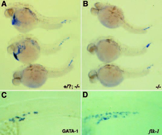
Fig. 7 GATA-1 is expressed in clo mutant embryos only in the region outlined by the flk-1-expressing cells. GATA-1 expression in wild-type and clo mutant embryos at 30 hpf. (A) Lateral view of 2 wild-type (top) and one clo mutant (bottom) embryos. (B) clo mutant embryos. (C,D) Higher magnification view of cells expressing GATA-1 (C) and flk-1 (D) in the lower trunk and tail regions of clo mutant embryos. GATA-1 expression is not detected at early stages in clo mutant embryos. Expression appears as flk-1 is being detected and only in the region lined by the flk-1-positive cells. Additional analysis reveals that although GATA-2 does not seem to be expressed in clo hematopoietic tissues, the GATA-1- expressing cells appear to mature normally as assessed by positive diaminofluorene staining used to detect heme (data not included).