
|
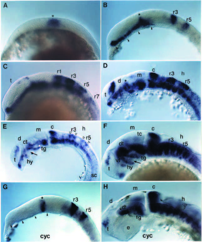
Fig. 2 In situ hybridization analysis of zp-50 expression during embryogenesis. All pictures are side views with the anterior part of the embryos oriented towards the left. In some cases the yolk has been removed for better visibility. Wild-type (A-F) and cyclops mutant (G,H) embryos were hybridized with a zp-50 probe. (A) bud (10 hpf). The asterisk marks the earliest expression domain in the forming posterior diencephalon (dorsal thalamus). (B) 5 somites (12 hpf). Rhombomeres r3 and r5 are indicated, as well as the early transverse zp-50 expression domain in the diencephalon (asterisk). The longitudinal expression domain in the ventral forebrain (arrowheads) is absent in cyc-/- embryos (cf. G). (C) 10 somites (14.5 hpf). Alternating high levels of zp-50 expression are found in odd-numbered rhombomeres r1, r3, r5 and r7. Expression begins in the telencephalon. (D) 20 somites (19 hpf). The asterisk marks the initial zp-50 expression domain in the dorsal thalamus. The initially contiguous longitudinal expression along the forebrain is subdivided into several domains. (E) 26 hpf. Open triangles indicate the lateral arches in rhombomeres r3 and r5. The lower part of these arches coincide with rhombomere boundaries. The arrow points to the forming ventral flexure. (F) 34 hpf. Hindbrain expression is observed in paired stripes along the rhombomere boundaries. (G) 5 somite cyc-/- embryo. zp-50 is expressed in rhombomeres r3 and r5 and the transverse domain in the posterior diencephalon (asterisk). Arrowheads indicate missing zp-50 expression in the ventroanterior brain. (H) 30 hpf cyc-/- embryo. Diencephalic zp-50 expression is found only in the dorsal thalamus in the area derived from the initial expression site visible after the bud stage (asterisk). c, cerebellum; d, diencephalon; dt, dorsal thalamus; e, eye; h, hindbrain; hy, hypothalamus; m, mesencephalon; sc, spinal cord; t, telencephalon; tc, tectum; tg, tegmentum; vt, ventral thalamus.