
Image
|
Figure Caption
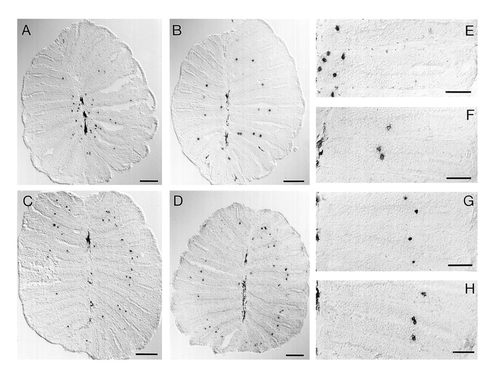
Fig. 3 Spatial expression patterns of odorant receptor molecules. Horizontal tissue sections of fresh-frozen nasal epithelium were hybridized with digoxigenylated riboprobes of fZOR6 (A and E), fZOR9 (B and F), fZOR8 (C and G), and fZOR5 (D and H). For orientation, compare Fig. 1. In A–D, the complete sections are depicted (cf. Fig. 1B). (Bar = 100 μm.) (E–H) Photomicrographs taken at higher magnification. (Bar = 50 μm.) Melanophores in the median raphe are visible as elongated dark stain (left edge of micrograph). Labeled cells are small, roundish, and dark.
Figure Data
Acknowledgments
This image is the copyrighted work of the attributed author or publisher, and
ZFIN has permission only to display this image to its users.
Additional permissions should be obtained from the applicable author or publisher of the image.
Full text @ Proc. Natl. Acad. Sci. USA