
|
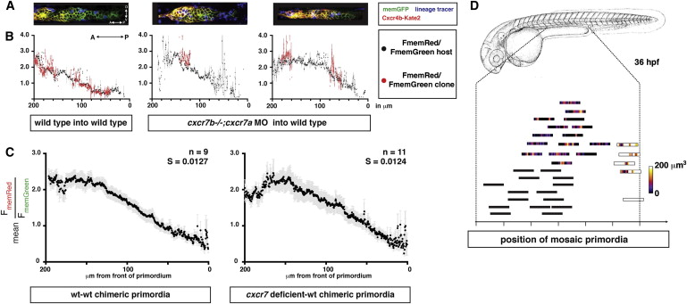
Fig. 5
Cxcr7 Modifies the Sdf1-Signaling Gradient across the Primordium at the Tissue Level
(A) Single confocal slices through mosaic primordia in live 36 hpf embryos of the indicated genotypes.
(B) Quantification of mean FmemRed/FmemGreen of host cells (black dots, gray bars SEM) and donor cells (red dots, light-red bars SEM) across the anterior-posterior axis of primordia shown in (A).
(C) FmemRed/FmemGreen ratio on the host cells only across wild-type-wild-type and cxcr7 deficient-wild-type chimeric primordia containing the Sdf1-signaling sensor. The front of the primordium is at 0 μm. Gray bars indicate SEM.
(D) Position of mosaic primordia compared to cxcr7b mutant (black rectangles) and wild-type primordia (white rectangle). The amount (heat map in μm3) and position of clonal tissue across 150 μm from the front of the schematized primordia is indicated.
Reprinted from Cell, 155(3), Venkiteswaran, G., Lewellis, S.W., Wang, J., Reynolds, E., Nicholson, C., and Knaut, H., Generation and Dynamics of an Endogenous, Self-Generated Signaling Gradient across a Migrating Tissue, 674-687, Copyright (2013) with permission from Elsevier. Full text @ Cell