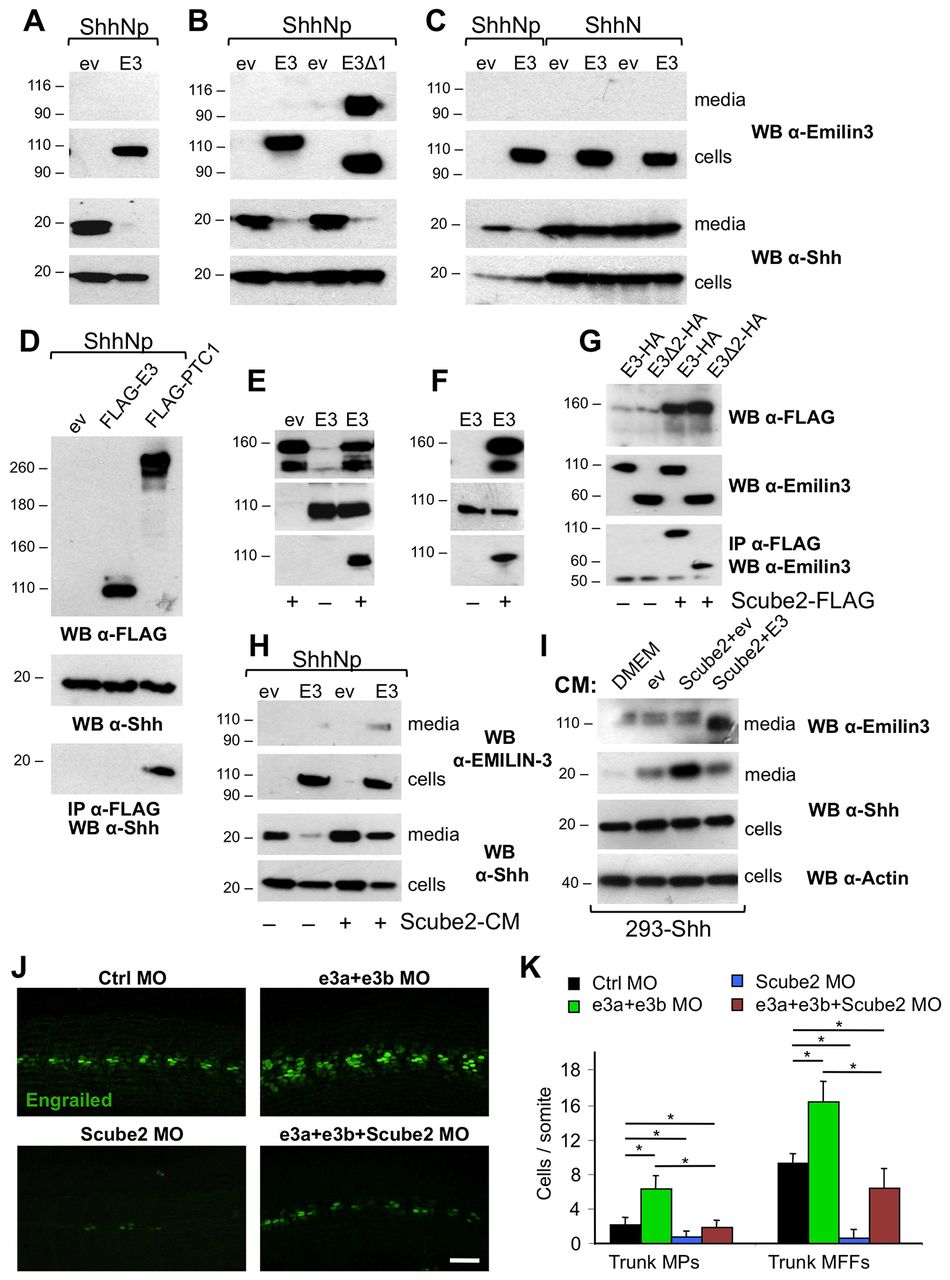
Fig. 4 Emilin3 functionally interacts with Scube2. (A-C) HEK293T were transiently co-transfected with the indicated constructs. Conditioned serum-free media and cell lysates were analyzed by western blot. (D) Cell lysates of HEK293T transfected with the indicated plasmids were analyzed by western blot or subjected to immunoprecipitation. (E-G) HEK293T were either co-transfected (E,G) or separately transfected (F) with the indicated plasmids. Media (E,F) and cell lysates (G) were analyzed by western blot or subjected to immunoprecipitation. (H) Transfected HEK293T were incubated for 24 hours with conditioned media derived from control or Scube2-transfected cells. (I) Stably transfected 293-Shh cells were incubated for 6 hours with fresh medium (DMEM) or with conditioned media derived from HEK293T co-transfected with the indicated plasmids. Media and cell lysates were analyzed by western blot. (J) Lateral views of 24 hpf embryos injected with the indicated morpholinos and immunostained for engrailed. Scale bar: 50 μm. (K) Quantification of engrailed-positive cells in injected embryos (*P<0.05; n=6). Data are mean+s.e.m. CM, conditioned media; E3, murine full-length Emilin3; E3Δ1, murine Emilin3 lacking the EMI domain; E3Δ2, murine Emilin3 lacking the EMI domain and the coiled-coil region; ev, empty vector; IP, immunoprecipitation; PTC1, human FLAG-patched-1; WB, western blot; Ctrl MO, control morpholino; e3a+e3b MO, emilin3a + emilin3b morpholinos; Scube2 MO, Scube2 morpholino; MPs, muscle pioneers; MFFs, medial fast fibers.
Image
Figure Caption
Figure Data
Acknowledgments
This image is the copyrighted work of the attributed author or publisher, and
ZFIN has permission only to display this image to its users.
Additional permissions should be obtained from the applicable author or publisher of the image.
Full text @ Development

