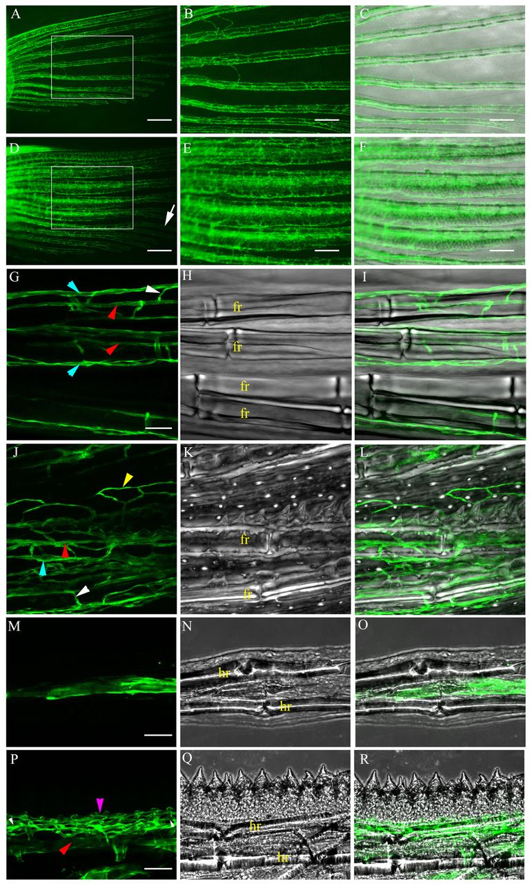
Fig. 2 Vasculature in Tg(fli1a:EGFP) pectoral fins. (A-C) Blood vessels (EGFP, green) in female zebrafish. (D-F) BT clusters in males are highly vascularized (boxed in D), but not in surrounding areas (arrow in D). (B,C,E,F) Higher magnification of the boxed regions in A,D. (C,F) Merged brightfield and fluorescent images. (G-R) Confocal micrographs. (G-I) Female with a single artery (red arrowheads), two veins (blue arrowheads) and intervessel commissures (white arrowhead). (J-L) Male with vascularized BTs, an artery (red arrowhead), intervessel commissures (white arrowhead), interray vessels (yellow arrowhead) and veins (blue arrowhead). (M-R) Longitudinal sections. (M-O) Female showing blood vessels that lie between the two hemirays. (P-R) Male showing blood vessels present in a web-like pattern (pink arrowhead) in the epidermis and above the artery (red arrowhead). fr, fin ray; hr, hemiray. Scale bars: 200 μm in A,D; 100 μm in B,C,E,F; 50 μm in G-L; 25 μm in M-O; 50 μm in P-R.
Image
Figure Caption
Acknowledgments
This image is the copyrighted work of the attributed author or publisher, and
ZFIN has permission only to display this image to its users.
Additional permissions should be obtained from the applicable author or publisher of the image.
Full text @ Development

