
|
Fig. 6
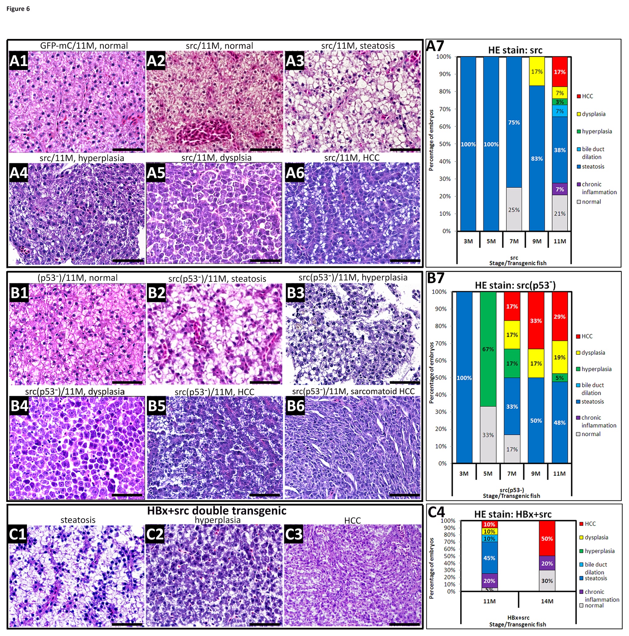
(A1) H&E staining of liver sections from wild-type fish revealed normal histology at 11 months. (A2~A6) H&E staining of liver sections from src-overexpressing, wild-type fish displayed steatosis, hyperplasia, dysplasia and HCC at 11 months. (B1) H&E staining of liver sections from the p53 mutant fish showed normal features at 11 months. (B2~B6) H&E staining of liver sections from src(p53-) transgenic fish showed severe steatosis and chronic inflammation, dysplasia, HCC and sarcomatoid HCC at 11 months. (C) The hepatocytes from the double transgenic line overexpressing HBx and src in a wild-type background exhibit steatosis, chronic inflammation, hyperplasia, and HCC. All sections were stained with H&E and photographed at 400X magnification. Scale bars: 50 μm. A7, B7, and C4 show the statistical analysis of the H&E staining results. The following different colors denote the different pathological features: gray-normal, purple-chronic inflammation, blue-steatosis, light blue-bile duct dilation, green-hyperplasia, yellow-dysplasia, and red-HCC.