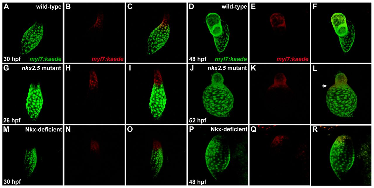
Fig. 9 Ventricular-to-atrial transformation occurs in the absence of Nkx gene function. Confocal projections of live zebrafish hearts carrying Tg(myl7:kaede) are depicted immediately following photoconversion at 26-30 hpf (A-C,G-I,M-O) and after later visualization at 48-52 hpf (D-F,J-L,P-R); arterial pole to the top. Green fluorescence of Kaede (A,G,M) is converted to red fluorescence (B,H,N) following UV exposure of the region of interest; merged views (C,I,O) indicate that labeled cells are at the arterial pole of the heart tube. At later time points, retained red fluorescence (E,K,Q) is visualized together with the green Kaede (D,J,P) that is continually produced by the transgene; merged views (F,L,R) indicate the locations of the labeled cells. (A-F) In wild-type embryos, labeling of ventricular cells near the arterial pole of the heart tube (A-C) yields labeled cells within the ventricular chamber at 48 hpf (D-F) (n=17/17). (G-L) By contrast, comparable photoconversion of ventricular cardiomyocytes at the arterial pole of the nkx2.5 mutant heart tube (G-I) generates labeled cells both within the diminutive ventricular chamber and expanding into the atrial chamber (J-L, arrowhead in L) (n=10/12). (M-R) In Nkx-deficient embryos, labeled cells from the vmhc-expressing portion (supplementary materialFig. S9N) of the heart tube (M-O) are later detected within the single, amhc-expressing (supplementary material Fig. S9P) chamber of the heart (P-R) (n=2/2).
Image
Figure Caption
Figure Data
Acknowledgments
This image is the copyrighted work of the attributed author or publisher, and
ZFIN has permission only to display this image to its users.
Additional permissions should be obtained from the applicable author or publisher of the image.
Full text @ Development

