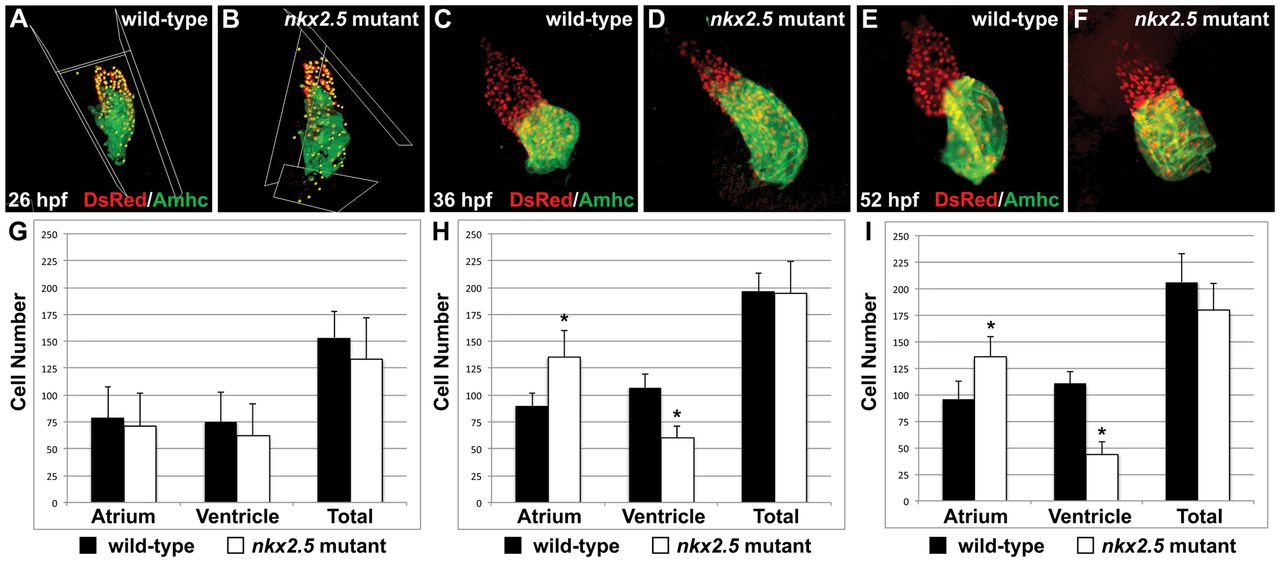
Fig. 2 Increased atrial and decreased ventricular cell numbers are first evident in nkx2.5 mutants following heart tube extension. (A-F) Immunofluorescence indicates expression of the transgene Tg(-5.1myl7:nDsRed2) (red) in both cardiac chambers facilitating cardiomyocyte counting at 26 hpf (A,B), 36 hpf (C,D) and 52 hpf (E,F). Atria are labeled with the anti-Amhc antibody S46 (green). (G-I) Bar graphs indicate numbers of atrial and ventricular cardiomyocyte nuclei, as well as the total number of cardiomyocytes; mean and s.e.m. of each data set are shown, and asterisks indicate statistically significant differences from wild type (P<0.001). (G) At 26 hpf, we find no statistically significant difference in cell numbers in wild-type (n=20) and nkx2.5 mutant (n=7) embryos. (H) At 36 hpf, comparison of wild-type (n=16) and nkx2.5 mutant (n=10) embryos reveals an increase in atrial cell number and a decrease in ventricular cell number in nkx2.5 mutants. (I) At 52 hpf, comparison of wild-type (n=16) and nkx2.5 mutant (n=11) embryos reveals an increase in atrial cell number and a decrease in ventricular cell number in nkx2.5 mutants.
Image
Figure Caption
Figure Data
Acknowledgments
This image is the copyrighted work of the attributed author or publisher, and
ZFIN has permission only to display this image to its users.
Additional permissions should be obtained from the applicable author or publisher of the image.
Full text @ Development

