
|
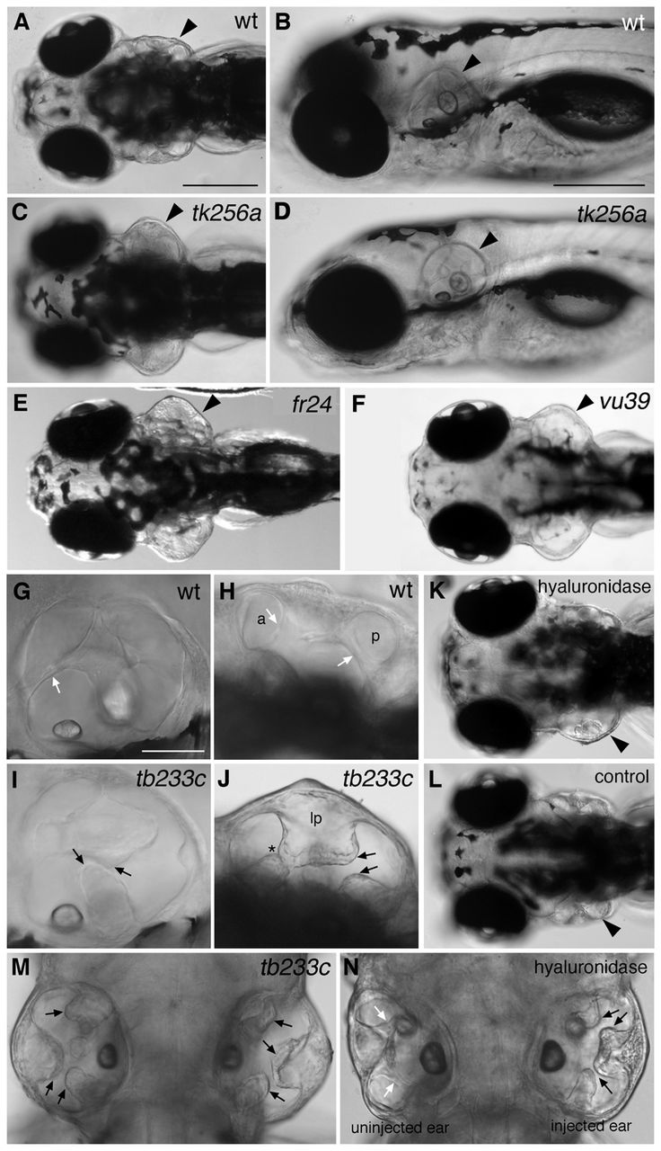
Fig. 2 lauscher mutant zebrafish have swollen ears at 5 dpf and defects in semicircular canal formation. (A-F) Live images of wild-type sibling (A,B) and homozygous mutant (C-F) embryos at 5 dpf. The ears (arrowheads) of the mutants are swollen at 5 dpf, but otoliths appear normal. (G-J) Live images of wild-type sibling (G,H) and tb233c mutant (I,J) ears at 5 dpf. (K) Injection of hyaluronidase into the lateral projection of the left ear of a wild-type embryo results in the collapse of the projection, failure of fusion and a swollen ear. (L) Injection of PBS into the lateral projection of the left ear has no effect on ear development. Arrowheads mark the injected ears. (M,N) Comparison of ear swelling and projection fusion in the tb233c allele (M) with an ear in which the lateral projection has been injected with hyaluronidase (N) (dorsal views). Black arrows mark unfused projections; white arrows mark fused pillars. Asterisk in J marks projections that have touched but not fused correctly. Abbreviations: a and p, lumens of anterior and posterior semicircular canals; lp, enlarged lateral projection. A,C,E,F,H,J-N are dorsal views. Scale bars: in A, 200 μm for A,C,E,F,K,L; in B, 200 μm for B,D; in G, 50 μm for G-J.