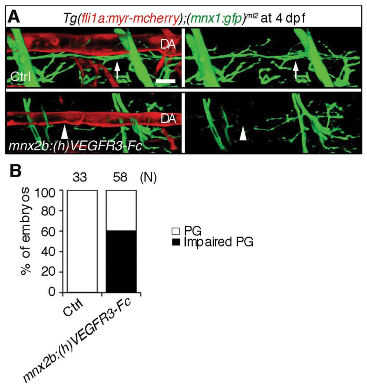
Fig. 7 Inhibition of Vegfr3 in the motoneuron results in impairment of alignment of the motoneuron axons and dorsal aorta. (A) 3D-rendered confocal stack images (lateral view) of Tg(fli1a:myr-mcherry);(mnx1:gfp)ml2 embryos (upper panel) and those transiently expressing (h)VEGFR3-Fc in the motoneurons under the control of the mnx2b promoter by Tol2-mediated gene transfer (lower panels). Left panels, merged images of mCherry and GFP; right panels, GFP images. Arrows indicate the motoneuron axon beneath the dorsal aorta (DA). Arrowheads indicate the impairment of parallel growth of motoneuron axon with the DA. Scale bar: 25 μm. (B) Quantitative analyses of the impaired parallel growth (PG) of the embryos grouped as in A. The number (n) of the embryos observed indicated at the top.
Image
Figure Caption
Acknowledgments
This image is the copyrighted work of the attributed author or publisher, and
ZFIN has permission only to display this image to its users.
Additional permissions should be obtained from the applicable author or publisher of the image.
Full text @ Development

