Fig. 5
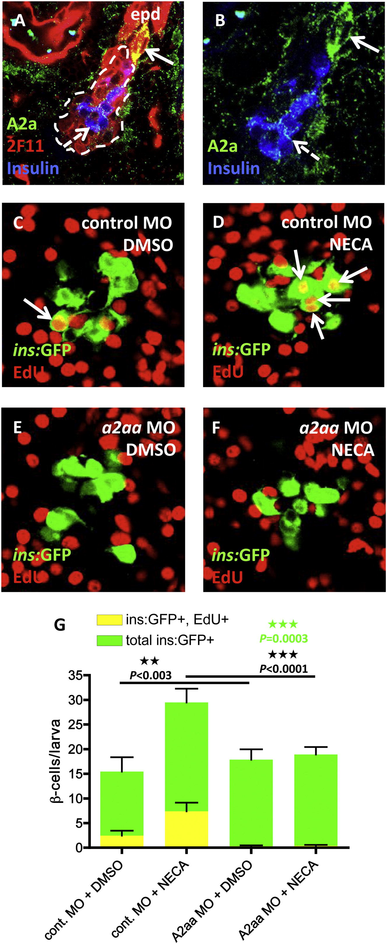
The Adenosine Receptor A2aa Mediates the Regenerative Effect of NECA
(A) Confocal image of the expression of the A2a adenosine receptors in a 5 dpf larva. The 2F11 antibody marks the extrapancreatic duct (epd) as well as the endocrine islet (outlined by the dashed line). High expression of A2a is found in cells budding off the epd (arrow), and low expression is found in insulin-expressing β cells (dashed arrow) and cells scattered throughout the exocrine pancreas.
(B) For clarity, a magnified view of (A), without the red color, is displayed.
(C–G) Tg(ins:GFP);Tg(ins:CFP-NTR) embryos were injected with a control MO or an a2aa MO at the one-cell stage and subsequently treated with MTZ from 3-4 dpf to ablate the β cells, and DMSO/NECA and EdU during β cell regeneration from 4-6 dpf. (C) Confocal image of a DMSO-treated control MO-injected larva displaying one β cell that had incorporated EdU (arrow). (D) Confocal image of a NECA-treated control MO-injected larva displaying four β cells that had incorporated EdU (arrows). (E) Confocal image of a DMSO-treated a2aa MO-injected larva where no β cells had incorporated EdU. (F) Confocal image of a NECA-treated a2aa MO-injected larva where no β cells had incorporated EdU. (G) Quantification of the total number of β cells and the number of β cells that incorporated EdU per larva during DMSO or NECA treatment of control MO-injected or a2aa MO-injected embryos. p values in black refer to Tg(ins:GFP)+, EdU+ cells, whereas the p value in green refers to total number of Tg(ins:GFP)+ cells. n = 11–25 larvae per group. Error bars represent SEM. See also Figure S6.
Reprinted from Cell Metabolism, 15(6), Andersson, O., Adams, B.A., Yoo, D., Ellis, G.C., Gut, P., Anderson, R.M., German, M.S., and Stainier, D.Y., Adenosine Signaling Promotes Regeneration of Pancreatic beta Cells In Vivo, 885-894, Copyright (2012) with permission from Elsevier. Full text @ Cell Metab.

