
|
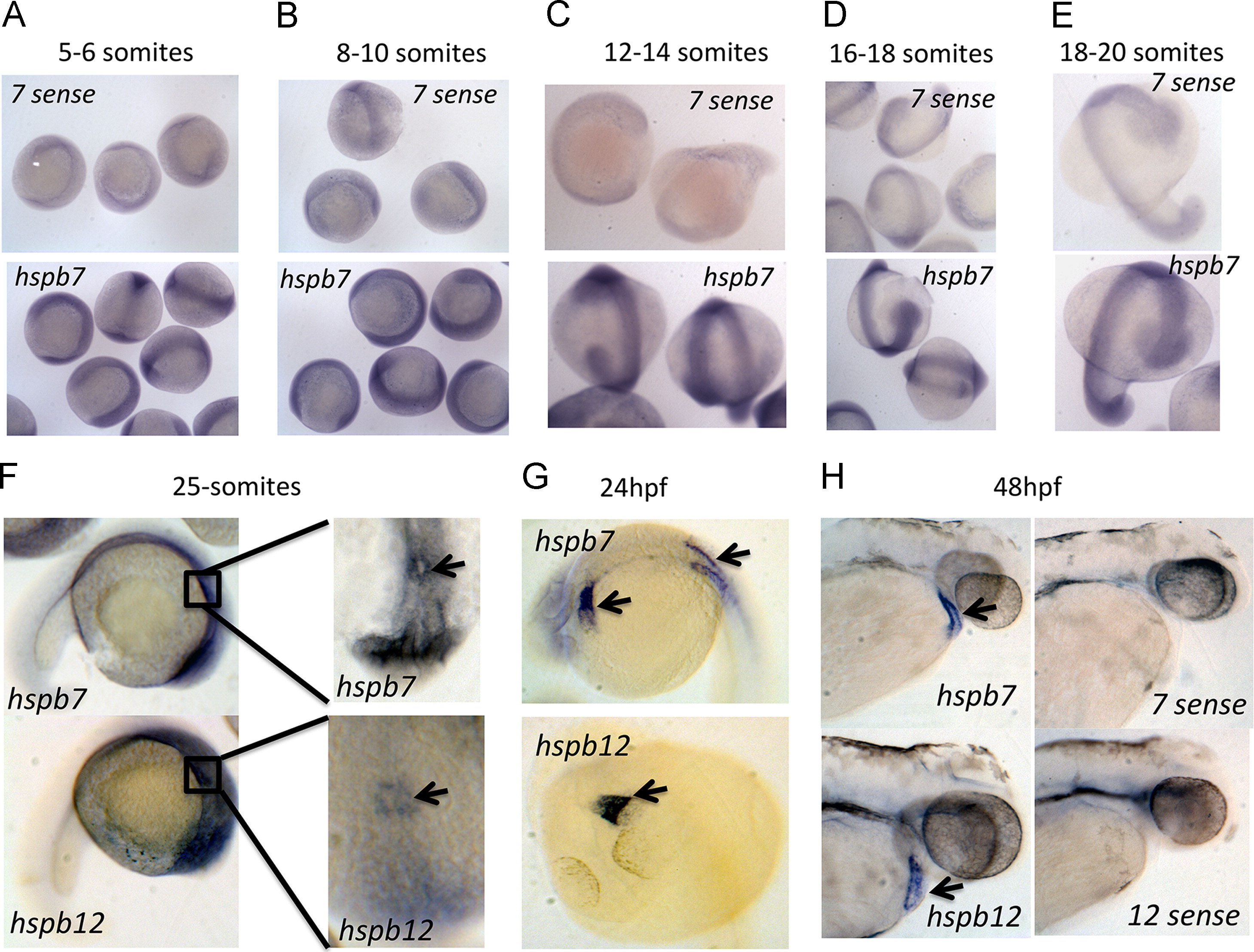
Fig. 1
Expression of hspb7 and hspb12 during zebrafish embryogenesis. (A–E) In situ hybridization expression data for hspb7 at 5–6 somites, 8–10 somites, 12–14 somites, 16–18 somites, and 18–20 somites, as indicated. Upper panels (7-sense) are controls at each time points for comparison to anti-sense (hspb7), demonstrating ubiquitous staining pattern between 5 and 20 somites. (F–H) In situ hybridization expression data for hspb7 and hspb12 at 25-somites, 24 h post fertilization, and 48 h post fertilization, as indicated. Insets in right panels of (F) show dorsal views of boxed regions in the lateral views of the left panels. Arrows indicate areas of expression including the cardiac cone (F), heart tube and somites (G), and heart (H).
Reprinted from Developmental Biology, 381(2), Rosenfeld, G.E., Mercer, E.J., Mason, C.E., and Evans, T., Small heat shock proteins Hspb7 and Hspb12 regulate early steps of cardiac morphogenesis, 389-400, Copyright (2013) with permission from Elsevier. Full text @ Dev. Biol.