
|
Fig. S9
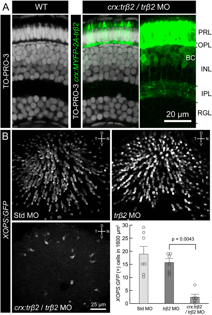
Ectopic expression trβ2 driven by the crx promoter decreases the density of rods. (A) trβ2 morphants rescued by trβ2 expression under the crx promoter (crx:trβ2/trβ2 MO) did not affect the laminar structure of the retina. Cell nuclei were stained with TO-PRO-3 (Invitrogen) in a wild-type retina (Left) and crx:trβ2/trβ2 MO retina (Center). (Right) Bipolar cells express trβ2 in crx:trβ2/trβ2 MO, but their axonal projections were not affected. MYFP signal in photoreceptors is saturated to demonstrate faint MYFP fluorescence in bipolar cell axons. INL, inner nuclear layer; IPL, inner plexiform layer; OPL, outer plexiform layer; PRL, photoreceptor layer; RGL, retinal ganglion cell layer. (B) Comparison of XOPS:GFP expression at 5 dpf between GeneTools standard control morphant (Std MO), trβ2 morphant, and crx:trβ2/trβ2 MO. The graph represents quantification of rod density (mean ± SEM). Circles indicate data from individual eyes. Statistical significance was determined using the Mann–Whitney rank-sum test. It is likely that the crx promoter used in this study is also active in rod precursors. The reduction of rods in crx:trβ2/trβ2 MO may be attributable to cell death of rod precursors or rods. Thyroid hormone induces cell death during metamorphosis in amphibians (16). Alternatively, a change in cell fate may have occurred because the density of photoreceptors increased in the “rescue” animal (see the figure legend of Fig. 4).