Image
Figure Caption
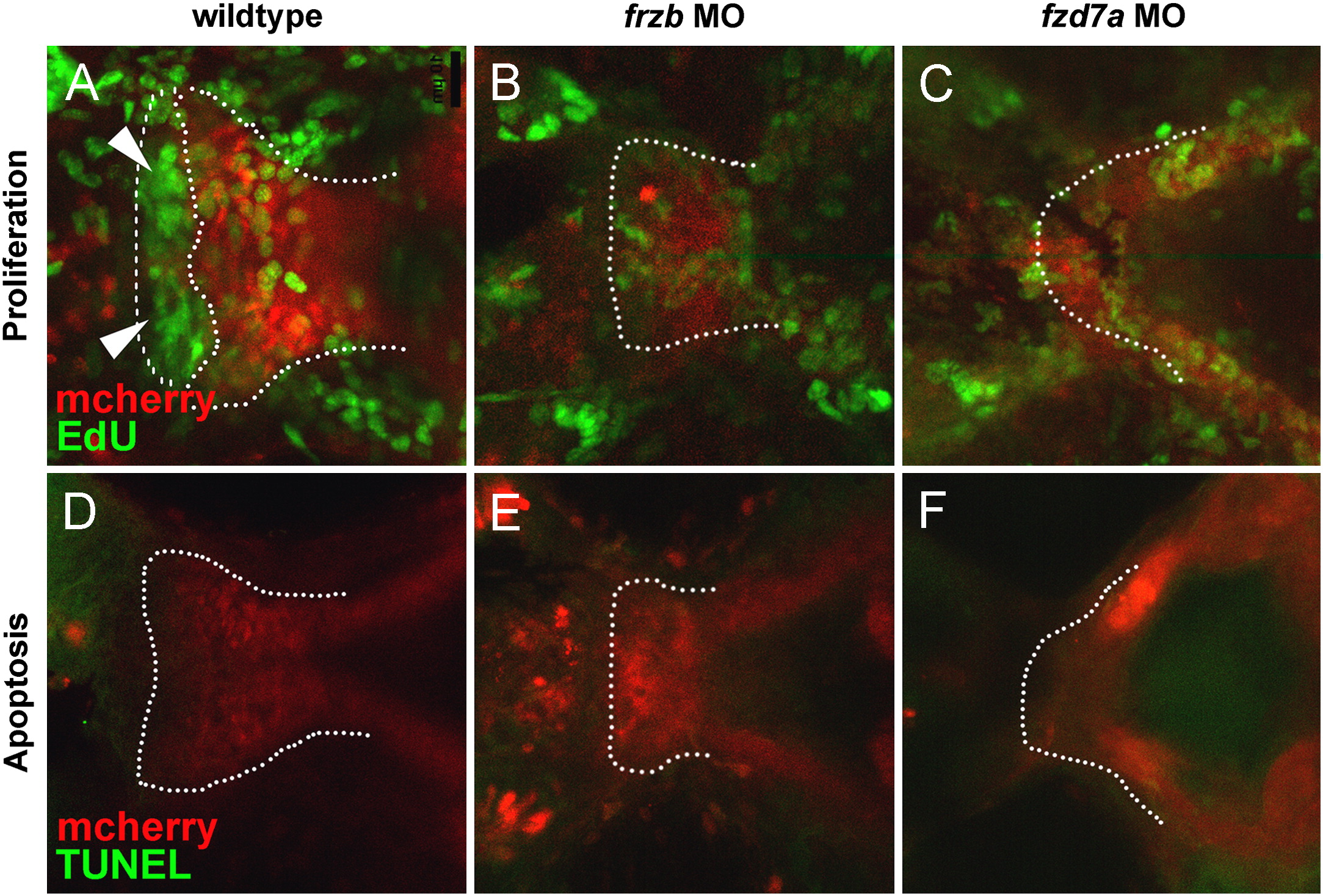
Fig. 5 frzb and fzd7a are required for chondrocyte proliferation. Anterior is to the left and all images are in ventral view. (A–F) EdU staining for new cell division from 55–60 hpf, a period of early maxillary extension, revealed a proliferative front of chondrocytes at the leading edge of the developing palate during normal development (A, arrows, dotted line) and the paucity of new cell division during this same period in frzb (B) and fzd7a morphants (C). TUNEL assay for apoptosis demonstrated that the shortened palate is not a result of increased cell death (D–F).
Figure Data
Acknowledgments
This image is the copyrighted work of the attributed author or publisher, and
ZFIN has permission only to display this image to its users.
Additional permissions should be obtained from the applicable author or publisher of the image.
Reprinted from Developmental Biology, 381(2), Kamel, G., Hoyos, T., Rochard, L., Dougherty, M., Tse, W., Shubinets, V., Grimaldi, M., and Liao, E.C., Requirement for frzb and fzd7a in cranial neural crest convergence and extension mechanisms during zebrafish palate and jaw morphogenesis, 423-33, Copyright (2013) with permission from Elsevier. Full text @ Dev. Biol.

